This is displayed by LPFT – what is a just culture exactly? This is completely ridiculous and biased. Just mention a “just culture” to all the parents and carers affected right now and particularly all the patients stuck on wards for years on end whilst Trusts such as this act dishonestly. When you dare to challenge you can expect a response as follows from the likes of Ms Munro who is as unfit to write complaint responses as the doctors who carried out the flawed capacity assessments.
Here is what Elizabeth has described her experience as: “hell on earth”. This is also how parents and carers view matters, those who are affected and those who are ignored. How can there be any accountability whilst those at the top have connections with the Care Qualilty Commission? How can any members of the public have faith in the Care Quality Commission who have produced some very dismissive responses even calling themselves a “business”. I have featured this on a former blog.
The Care Quality Commission (CQC) have asked us to respond to requests you have raised with them and clarify the Trust’s position on a number of points:
You have requested an apology for the verbal threats made by Dr Khokhar where she said “I am banning you indefinitely for inciting your daughter to attack members of staff on Xmas Day”.
Following the incident on Christmas Day 2023, the Trust followed the usual incident reporting and investigation processes, as well as complying with instructions and communication from the Police. During this period of investigation, your visits to the ward to see Elizabeth were suspended in line with the Mental Health Act Code of Practice 2015 and to ensure the effective delivery of Elizabeth’s care and treatment could be prioritised. Dr Khokhar accepts that he used the word “indefinitely” and that this could be seen as inflammatory. It was used to reflect that at the point, an end date to the restrictions could not be confirmed. However, the letter sent dated 29th December 2023 confirmed that the situation would be reviewed on 25th January 2024. After subsequent review of the relevant documentation, the visiting restrictions were lifted from the 5th February 2024.
You asked about your invoice for £50 for Elizabeth’s ‘wasted’ phone contract.
As previously explained in an email on 29th Februay 2024, Elizabeth’s phone access was limited for a fixed period of time as part of a comprehensive care plan that considered Elizabeth’s human rights. This was to ensure the response to her phone use did not adversely impact her engagement with, or the efficacy of, her treatment plan. This ceased on the 5th February 2024 following a review of all of the relevant factors. Elizabeth’s phone continues to be kept securely by ward staff and Elizabeth is able to request to use her phone at any time. In addition, ward staff regularly encourage Elizabeth to use her phone to maintain contact with family.
We monitor and document Elizabeth’s phone use to ensure that she has fair and reasonable access to her phone. This details the occasions when she has been supported to use her phone and make calls and evidences that Elizabeth continues to be able to use her phone on request . On the basis that Elizabeth does continue to use her phone, we would not provide any reimbursement for her phone contract.
You said that Elizabeth’s birthday cake was thrown away.
To ensure that the limited storage space for fresh food brought onto the ward by visitors is fairly available to all patients on the ward, a new process has been put in place. This is clearly communicated to patients and their friends and relatives when they visit to manage expectations. The ward also adheres to Trust wide infection prevention and control (IPC) policies relating to food storage and consumption to ensure patient safety. Elizabeth’s birthday cake did have to be disposed of on 15th February 2024 as the expiry date had been exceeded.
You raise concerns about the size of the visitor’s room and that it was cramped which upset Elizabeth
The family room on the ward is used for visits as it affords privacy and has CCTV available which can be used to review the area, if necessary. This room is the largest room on Castle Ward used for visitors.
Subject Access Requests
In relation to the Subject Access Requests (SARs) received both for personal data relating to yourself and requesting a copy of Elizabeth’s rapid tranquilisation healthcare records, the Trust’s position remains the same as detailed in the email sent on 11th December 2023. This is because we are of the view that your request is “manifestly unfounded or excessive” as detailed in Article 12 (5) of the GDPR.
Our Records Management team did note that you subsequently attached a consent form from Elizabeth. However, this was not the correct consent form to support your Subject Access Request. Further, we continue to have concerns regarding Elizabeth’s capacity to consent, which has historically remained interchangeable with regards to information sharing. We also have continued concerns that any information shared may, in turn end up within a public forum which is outside the scope of any consent that we may gain.
We ;have provided a copy of this letter to the CQC.
Yours sincerely
Ann Munro
Patient Experience Lead
Cc Care Quality Commission (Central) Citygate, Gallowgate, Newcastle Upon Tyne
NE1 4PA
EXTRACTS FROM MY RESPONSE EMAIL TO MS MUNRO’S LETTER.
Yr Ref MHA-011217-X6F7T0
Dear Ms Munro
CEO (Sarah Connery) on the Trust’s Executive Board also works/worked as a Specialist Advisor for CQC or The Director of Operations (Chris Higgins) who works/has worked as an Executive Reviewer for CQC
Thank you for your letter of 4 April.
When you mention “The Care Quality Commission” asked you to respond. Who might that be? Could that be either of two LPFT Board Executives mentioned above by any chance? To think, all this time so it would appear LPFT have been answering on behalf of the Care Quality Commission in a defensive, almost threatening manner. What a conflict of interest! No wonder why none of my complaints have been dealt with satisfactorily and with Mr Higgins as Executive Reviewer for CQC it is also no wonder why you are rated “good”.
How can you have a CEO (Sarah Connery) on the Trust’s Executive Board who also works/worked as a Specialist Advisor for CQC or The Director of Operations (Chris Higgins) who works/has worked as an Executive Reviewer for CQC however I am talking about LINCOLNSHIRE PARTNERSHIP TRUST WHO APPEAR TO HAVE THEIR OWN LAW TO SUIT THEMSELVES.
I would like your opinion on this conflict of interest, Ms Munro.
Regarding your third paragraph I would point out this was the third time Police were called unnecessarily, all done for the purpose of threatening and intimidation however Section 9 statements were done to back the truthfulness of events on Xmas Day to cover our backs. What a waste of Police time. It is obvious that the restrictions are still ongoing with HCA’s/nurses writing notes during visits (no doubt of an adverse nature). This is bullying to the extreme to stop visits and threaten to do so indefinitely but still continue with restrictions. You might like to write a statement t explaining exactly what you mean by “suspension in line with the MHA Code of Practice”: Please also explain why I am being treated differently to everyone else which is discrimination.
Here is the MHA Code of Practice relevant points which were not even quoted in full as a reason to ban my visits.
MHA CODE OF PRACTICE
Restrictions or Exclusion on Clinical Grounds
11.14
From time to time, a patient’s Responsible Clinician may decide, after assessment and discussion with the MDT, that some visits could be detrimental to the safety or wellbeing of the patient, the visitor, other patients or staff on the ward.
In these circumstances, the Responsible Clinician may make special arrangements for the visit, impose reasonable conditions or, if necessary, exclude the visitor. In any of these cases, the reasons for the restriction should be recorded and explained to the patient and the visitor, both orally and in writing (subject to the normal considerations of patient confidentiality). Wherever possible 24 hour notice should be given of this decision.
Restriction Or Exclusion on Security Grounds
11.15
The behaviour of a particular visitor may be disruptive in the past, to the degree that exclusion from the hospital is necessary as a last resort. Examples of such behaviour include:
Incitement to abscond
Smuggling of illicit drugs or alcohol into the hospital or unit
Transfer of potential weapons
Unacceptable aggression
Attempts by members of the media to gain unauthorised access
11.16
A decision to exclude a visitor on the grounds of their behaviour should be fully documented and explained to the patient orally and in writing.
Where possible and appropriate the reason for the decision should be communicated to the person being excluded (subject to the normal considerations of patient confidentiality and any overriding security concerns.
LPFT ARE IN BREACH OF THE MHA CODE OF CONDUCT RE: THE FOLLOWING POINTS:
“may make special arrangements for the visit” : No special arrangements were ever made and contact severely restricted by taking the phone away that affected the entire family and was detrimental to Elizabeth – depriving her of her phone deprived her of contact with everyone else in the family and was done primarily to punish me and deprived Elizabeth of listening to her music. This is in breach of human rights and totally degrading and undignified to Elizabeth..
“the reasons for the restriction should be recorded and explained to the patient and the visitor, both orally and in writing”.: None of this was done properly explaining the grounds mentioned below for banning visitors. No Impact assessment carried out as should have been.
“subject to the normal considerations of patient confidentiality). Wherever possible 24 hour notice should be given of this decision” This was not done. The pre-arranged visit was cancelled on the spot whilst Elizabeth stood the other side of the door – absolutely degrading and totally cruelty to do this without any notice. Nursing staff acting ultra vires upon orders at Executive level and against their own code of conduct.
None of the above was done properly and also LPFT failed to carry out an Impact Assessment which LPFT is supposed to do under NHS Guidelines and this should have been done on myself, on Elizabeth and a copy sent to the Nearest Relative. LPFT are therefore in breach of NHS Guidelines. Here below in bold are the justified reasons to completely ban visitors according to MHA Code of Conduct.
Incitement to abscond
smuggling of illicit dugs or alcohol into the hospital or unit
Transfer of potential weapons
Unacceptable aggression
Attempts by members of the media to gain unauthorised access
None of the above apply in my case.
Dr K cannot very well deny threatening me with an indefinite ban as this was witnessed. Having such a ban has had a detrimental affect on my health. Elizabeth’s face is covered in marks where she is distressed and has been self harming and has said on more than one occasion she no longer wishes to live which in turn affects my health. The missing of meals has continued beyond the last safeguarding and had happened constantly at Ash Villa in the past yet nothing was done. It is bullying to the extreme to ban visitors and that is the tactic that LPFT adopted. My visits were suspended on none of the grounds above. Elizabeth’s care and treatment has been atrocious. She has been missing meals, isolating in her room because she cannot stand the noise. She has been deprived of proper pathological tests and nothing has been arranged as regards Sheffield as I have checked. There is no way a MH team can do proper pathological observations and have been filming my daughter when she is having fits against her wishes. This is an absolute disgrace. The way the MCA has been abused is another disgrace on the part of LPFT who enlisted the assistance of the council in this respect at County Court that lasted months and months on end. Human rights are non existent under LPFT and have never taken into account of. My daughter’s human rights are being dreadfully abused. It is not true when you state visiting restrictions were lifted from 5 February 2024. Restrictions are very much ongoing and visits are not of quality time but intrusive and degrading due to constant supervision. I am being discriminated against because other family members do not have 2-1 restrictive visiting with members of staff writing notes. The restrictions still very much in place clearly have a detrimental affect on my daughter who is treated like a prisoner. She reacted on Xmas Day to staff taking notes constantly. Even on Xmas Day, human rights were ignored by LPFT. How would any of you like this kind of treatment? It is breach of Art 8, Art 5 and Art 3 HRA. Police time was wasted on Xmas Day and Elizabeth was thrown into seclusion and rapidly tranquilised when she became upset by a crowd of “professionals” waiting outside the visitor’s room who grabbed hold of her because she had shouted at the lady writing notes because it was totally distressing to her.
There have been two accidents apparently according to Elizabeth, one at Ash Villa and another on Castle Ward where she lost balance or hit her head badly. Why wasn’t anyone in the family notified?
Because we pay a phone bill each month and Elizabeth hardly gets any use of the phone it is only fair that LPFT compensates for this. The bill now stands at £60 for your information and I request settlement of this amount to Elizabeth as already advised. Every month she is restricted with her phone I am adding on to that bill. Also under Ash Villa the phone was taken away for weeks on end making it difficult for the entire family to get through and for Elizabeth to be able to speak to family members. The reason for this was because Elizabeth kept calling the Police in distress. It says so in file papers.
“Elizabeth’s phone continues to be kept securely by ward staff and Elizabeth is able to request use of her phone at any time.” That is in breach of human rights. Totally degrading treatment – shame on you LPFT! Is this care? or rather is it abuse of a vulnerable person and their rights – it should be the least restrictive care under the MH LPFT are abusing patient’s rights and if this is just my daughter then this would also come under the Equality Act 2010. No other patient is being treated in such a degrading manner as my daughter. It is not the point that she is encouraged to use her phone. She is not at risk from anyone in her family and therefore the restrictions are excessive and the kind of thing that would apply to a restricted prisoner not a patient under the MHA.
LPFT haven’t got a clue when it comes to human rights and this is why I am copying in the British Institute of Human Rights. To keep the phone locked away constantly is a disgrace and still ongoing to date. Elizabeth does not like the phone being monitored and she expressly said she did not want to be filmed so why has this just gone ahead?
You clearly do not know the meaning of “fair and reasonable” . Elizabeth is a grown adult of 37 years of age and does not need support to use her phone.
The Birthday cake was not given to Elizabeth on her Birthday and should have been given to her immediately as I specially came back to the ward. It was her Birthday and no respect was given to this special occasion. Elizabeth did not even know I had brought her the Birthday cake so once again there was no communication. At Ash Villa all her presents were just dumped in a storage room. She did not know they existed. I brought another cake to make up for this and again Elizabeth was not given it. The second time it was not fresh cream and I assume thrown away.
Yes, the visitors room is cramped when 2 people are listening to every word and crammed into the room. It is undignified and unpleasant to say the least. How can you possibly say the visitor’s room affords privacy in the circumstances. You just have no insight into the situation. The rooms are all small in nature – not much difference in size but because of the restricted visiting and note taking going on (no doubt adversely against me), it is an unpleasant degrading experience that even a prisoner does not have to endure. In a prison, warders walk up and down but give respect to prisoners at visiting times to have privacy to see their families. The ward is worse than prison and the treatment is degrading, abusive and aimed no doubt as punishment towards me, but inflicting upon my vulnerable daughter.
Subject Access Requests.
I know LPFT do not want me to see any records especially not the copy of the CP11 rapid tranquilisation log. All the files and notes being taken will be requested through ICO. You have no right to refuse such request from both of us.
Your capacity assessments are totally flawed. I want full safeguarding done externally because in the past safeguarding under LPFT resulted in “I am satisfied” response. The safeguarding should be especially looking into the flawed capacity assessments. I am now a BI assessor – everything LPFT has done is wrong. Your capacity assessments are not fit for purpose. LPFT have abused the MCA. It is most certainly of public interest that everything is addressed openly and transparently.
Finally, please ensure that my daughter gets all the pathological tests she needs under Sheffield as soon as possible as this is another area of safeguarding that needs to be thoroughly looked into. I believe safeguarding should be done on the deprival of medical referrals to my daughter for her physical health and referrals need to be made to spend a full week under Sheffield for everything she needs, including Endocrinology
I have to say, I have never encountered such bullying in all my life and such dishonesty. You have been rating yourselves as good yet there are tremendous conflicts of interest I have witnessed at Executive level.
The capacity assessments are useless without referral to Sheffield and according to Professor Keith Brown everyone should be included (including myself) with the capacity assessments undertaken. My daughter most certainly has capacity and she is being abused and no-one cares otherwise this situation would not have been left to continue for so very long.
Here are the guidelines to filming patients against their wishes:
Keith Brown. Editor. Demystifying Mental Capacity: A guide for health and social care professionals (Post-Qualifying Social Work Practice Series). First …
Mental Capacity Act 2005 :
(Section 4): Requires that patient to be regarded as having capacity until evidence ascertained as to how that capacity is impaired.
(Section 4) (1) (a) and (b): “The Prohibited Step” Decision maker should not draw conclusion on capacity from patient’s age, appearance or on a condition of his/hers or an aspect of behaviour which might lead others to make unjustified assumptions on what might be in his/her best interests.
(Section 4) (2): Decision maker should try to identify all issues most relevant to the individual relating to particular decision (para 3, Main Code of Practice).
(Section D): inconsistencies of witness statements on capacity. Capacity assessments not conducted as required by S1 MCA 2005
Masterman-Lister v Brutton & Co [2003] 1WLR 151
“a party to legal proceedings is capable of understanding, with the assistance of such proper explanation (in broad terms and simple language) from legal advisers and other experts as the case may require, the matters on which their consent or decision was likely to be necessary in the course of those proceedings.”
Dont bother providing a copy to the CQC as LPFT are the CQC defending yourselves and rating yourselves as good when in actual fact you should be rated as “Requires improvement” or “inadequate” and you can’t even offer an apology for the shocking abuse of my daughter and her family in terms of human rights which you clearly have no insight of.
My final question to you is have LPFT applied for DoLs and in the meantime waiting to hear from the court because your intention is to dump her into a care home out of area in say Yorkshire and restrict contact forever? Why is she still held without leave under the MHA for over 2.5 years deprived of family contact and proper physical healthcare? I would like to hear from any of you in this respect as this is absolute abuse and breach of the law and the question needs to be asked about the validity of the entire section that is ongoing and the dishonesty in arriving at such decisions to hold her a prisoner indefinitely. My daughter is one of thousands trapped under the MHA by appalling Trusts such as LPFT and dishonesty too on the part of Councils who do not wish to provide any support whatsoever in the community. In fact I was not asking for anything of this area which brings me to the question of residency status and whether that responsibility still remains the responsibility of Enfield?
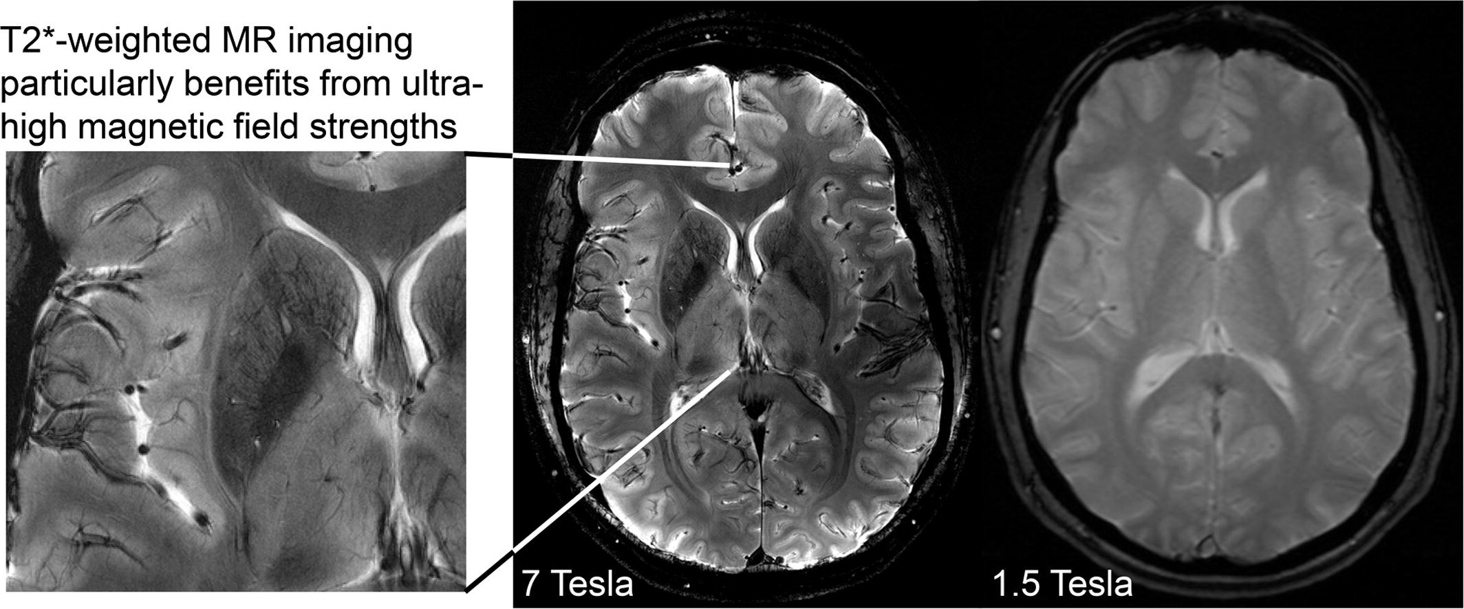
I have already revealed Elizabeth’s scans which were done in November at Lincoln County Hospital with the result of “normal”. I would urge all of my readers to double check what scanner has been used as a 1.5 scanner has not picked up what is clearly visible on the private scans I had done through S G Radiology. I am waiting for an explanation as to what the images portray as I cannot accept these images to be normal. They even state “trauma” so as a parent or carer it is evident that you cannot always assume that normal means normal. A while back Elizabeth had an ultrasound as there were fears of cancer. Now I want all the scans done again under a Tesla 3. I have heard a Tesla 7 is even better and will look into where this can be done in the UK. So Lincolnshire, Enfield, Weston are just three examples where they do not have the up to date scanner and therefore you cannot rely on the results. This affects not just people under MH but everyone.
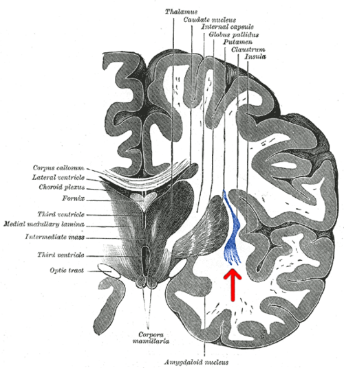
For so long now I have been trying to get answers – it clearly states “Anterior Region Medial Temperol Compromise which is not a mental illness and yet Elizabeth has been treated under MH with massive dosage of antipsychotics that do not work and is “treatment resistant” – poor/non metaboliser which I have prove by P450 tests. Now everything is falling into place and it is something I want to share with all of you especially those going through cancer treatment. Never trust the word “normal” and check what scanner your Trust has. I am currently asking my MP, Victoria Atkins why Lincolnshire does not have a scanner that is reliable. This means all of Elizabeth’s treatment may be affected as last year there was a cancer scare. Now I want all the scans/ultrasounds done through Sheffield and for Elizabeth to be referred to a Neurological Ward where for once her physical health can be properly assessed over a period of weeks. If the MH team stand in my daughter’s way of her physical health appointments and pathological tests I will report everything on here. I have already requested reports from all the professional bodies who have done nothing so far to stop the abuse to my daughter. Most importantly doctors should put the physical health of their patients first and foremost. I have now highlighted how patients are being deprived under just one area and how patients could have lost their lives due to the scans and how patients under the MH are refused pathological tests. This is why I was banned for months on end visiting my daughter and bullied by various doctors and this needs to be looked into. Police time has been wasted. Having lost close friends to cancer I intend to give this massive publicity.
LPFT under MH do not use PANSS??? Below is what I have written to Lincolnshire Partnership Trust as I am concerned for all the patients on Castle and Ellis Ward who may not have had reliable scans and I do not think anyone is being treated properly and wish to share so that everyone can be aware of the true facts:
PANSS
The PANSS score is an essential diagnostic tool which LPFT are not using and should be. All the scans may need to be re-done under scanners in Sheffield not just the MRI – everything and you may have to review all the patients on both Castle and Ellis Wards who have had scans done in Lincoln under the 1.5 Tesla in light of the revelations below.
The PANSS score is a long established test approved by the Royal College of Psychiatrists and used by NHS Mental Health Teams in many of the trusts to determine the severity of the schizophrenia the patient is suffering from. Its purpose is to confirm diagnosis, to guide the treatment regimen and to determine suitability for matters such as section 17 leave, suitability of the patient for post-discharge accommodation and eventual discharge from mental health care.
Here are LPFT’s comments below:
“LPFT does not use the PANSS scale for various reasons but does use the Glasgow Antipsychotic side effect scale (GASS). This measures the side effects of antipsychotics rather than the efficacy of antipsychotics”.
This is totally unsatisfactory for the following reasons. To begin with to quote LPFT’s own words “for various reasons” gives no explanation at all why this scale is not used by LPFT and is a cursory and unhelpful excuse.
The PANSS scale as mentioned above is crucial in determining not only treatment but deprivation of liberty and the ability of Elizabeth to enjoy leave and to have quality time off the ward. Elizabeth’s stifling detention without hope of even the chance to have a day out or to spend time with her family on important occasions like Christmas and her birthday are a contribution to her state of mind and a detriment to her eventual recovery.
The scale is also used to determine an appropriate treatment regimen which goes beyond the simple use or rapid tranquillisation and isolation preferred by the staff of the hospital in which she is detained. The hospital has obstructed all attempts to have Elizabeth’s mental health condition based on a determination that their ‘diagnosis ‘ of schizophrenia would be the only option. Elizabeth is now seeing a neurologist but that has only happened because that intervention was sought by me and was indeed actively discouraged and obstructed by the Responsible Clinician and the Clinical Lead on the ward.
The Glasgow Antipsychotic Side effect (GASS) scale that LPFT refer to in their email has an entirely different purpose to the PANSS scale and you are well aware of that. The GASS effect scale is for detecting adverse drug reactions and it has to be said if they are indeed using this they have ignored a number of these over the last two and a half years in spite of them being pointed out to them.
LPFT are fully aware that the GASS scale is not a substitute for PANSS but an entirely different test and the reply to the NR was disingenuous and unhelpful. To date no detailed explanation of why PANSS is not being used has been given and the Mental Health Act Team (notably the email does not identify its author) have failed to help once again. Please explain.
In the absence of proper monitoring by the people entrusted with Elizabeth’s care is it hardly surprising that after two and a half years subjected to a deprivation of liberty regime more restrictive than a section 37/41 order and constant prn rapid tranquillisation that she has made no improvement and is as far away from discharge as she ever has been.
The fact is the private scans are most certainly more superior to theirs and have been shown to numerous experts who are biomedical scientists yet they agree. If LPFT hadn’t spent a fortune getting rid of me as NR they would have more money to spend on decent scanners such as Tesla 3 and not having one is putting everyone’s life at risk not just those under MH. The taking away of my daughter’s right of capacity and autonomy is the biggest violation of all human rights. About 10 doctors apart from Dr Memons of Cygnet have stood in the way of her having MRI scans so that is why I was suspicious and arranged it myself. The MH system is ridiculous and I was the best NR and they got rid of me for their own convenience which again is abrogation of my daughter’s human rights. As highlighted in Medscape it is mentioned about brain tumours being missed and metastacising before being found which is potentially fatal which is why I am warning everyone not to trust their NHS scans and to check on which scanner has been used by their Trust.
It is also important to get brain lipids tested. They can affect both metabolising enzymes and result in ADRs. Several endocrine/metabolising disorders are associated with elevated levels of lipids (hyperlipidemia).
RESPONSIBLE CLINICIANS ARE SUPPOSED TO FOLLOW THE MHA 1983 BUT UNDER LPFT WHAT IS BEING DONE IS NOT CONDUCIVE TO ELIZABETH’S RECOVERY AND I WILL FEATURE MORE SHOCKING REVELATIONS ON MY NEXT BLOG. THEY NEEDED TO ROB HER OF HER CAPACITY SO THAT THEY COULD ABUSE THE CONCEPT OF THERAPEUTIC NECESSITY WHILST LABELLED WITH NO CAPACITY. I HAVE CAUGHT ON TO THEIR CRAFTINESS AS THIS GETS THEM OUT OF CONSULTING WITH HER OR RECORDING HER WISHES AND THIS IS WHY THEY GOT RID OF ME AS NR AND THIS IS TO PROTECT THEMSELVES. NO HUMAN RIGHTS AT ALL ARE TAKEN INTO ACCOUT BY LPFT. THE TERRIBLE DENIAL OF LIFE EXPERIENCE IS DETRIMENTAL TO MY DAUGHTER’S MENTAL AND PHYSICAL HEALTH AND TOTAL ABROGATION OF MEDICAL ETHICS. EVEN A S37/41 PATIENT IS NOT DENIED SUCH RIGHTS. HOW CAN HER DETENTION BE LAWFUL UNDER MHA 1983. THEY CONTINUALLY FLOUT THE STATUTORY SAFEGUARDS. THEY SHOULD BE SUBJECTED TO JUDICIAL REVIEW AND PROFESSIONAL MISCONDUCT ACTIONS.
Why 3T is necessary for detecting lesions
■ In more than 500 follow-up images, only four of 1996 new or enlarged multiple sclerosis lesions would have been missed with 3.0-T MRI without the administration of contrast material.
■ With 3.0-T MRI, the assessment of interval progression did not differ between contrast-enhanced and nonenhanced images.
Introduction
Inflammatory lesions in multiple sclerosis (MS) are detected as focal areas of high signal intensity on T2-weighted MR images. By depicting newly occurring lesions, MRI reveals subclinical disease activity. Therefore, regular follow-up MRI is considered a mainstay of clinical care for patients with MS or clinically isolated syndromes.
Earlier studies have reported that the administration of contrast material is necessary to maximize sensitivity for detecting new lesions. However, these results date back more than 2 decades and were based on two-dimensional images obtained with 4–5-mm-thick sections at magnetic field strengths of 1.5 T and lower.
MRI units with higher field strengths have become widely available, especially for brain imaging. In addition, three-dimensional isotropic MRI sequences were introduced and were shown to outperform conventional two-dimensional sequences in lesion depiction; they are therefore part of recommended MRI standards in MS . Furthermore, the double inversion-recovery (DIR) sequence was introduced. Although this sequence is best known for its ability to depict cortical lesions, it is also useful for depicting white matter lesions Recently, longitudinal subtraction techniques have been developed that show new or enlarged lesions as bright spots while pre-existing lesions and normal-appearing brain parenchyma are canceled out.* Such techniques substantially improve the sensitivity in the detection of new or enlarged lesions in MS at follow-up imaging.
We hypothesized that the use of contrast material does not improve sensitivity in the detection of new or enlarged lesions at follow-up MRI when modern three-dimensional sequences performed at a field strength of 3.0 T are used together with longitudinal subtraction maps. We therefore performed this study to investigate whether the use of contrast material has an effect on the detection of new or enlarged MS lesions and, consequently, the assessment of interval progression.
*The private 3T scans are much more able to see the lesions that the inappropriate 1.5T scanner cannot see.
The higher resolution scans cancel out the distortions and signal noise that hide the lesion and make the scan look normal.
1.5T MRI scanners are not fit for purpose in discovering brain lesions and inflammation.
Brain lesions and inflammation, especially in the temporal lobe are responsible for poor drug response and in some cases for diagnosis.
1.5T scans are responsible for false negative (normal) brain images.
Neuro Second Opinion Catches Missed Brain Lesions Suggestive of MS
American Roentgen Ray Society Dynamic contrast-enhanced MRI (DCE-MRI) has clearly been shown to be a highly sensitive tool for the detection of breast cancer [1–14]. Reported high sensitivity (83– 100%) [1–5] of MRI for breast cancer led im[1]agers initially to presume that non-enhancing legions on MRI were benign and did not warrant biopsy [1, 15]. However, subsequently reported articles have shown that all malignant lesions do not show enhancement at DCE[1]MRI [2, 5, 16–20], with enhancement absent in up to 12% of known malignant lesions. In a recent multi-institutional study of 995 lesions in 854 women, Schnall et al. [20] reported that 16% of 77 ductal carcinoma in situ (DCIS) lesions and 3% of 422 invasive cancers showed no enhancement. Teifke et al. [18] found 28 (8.4%) of 334 invasive cancers and 13 (65%) of 20 non-invasive cancers were missed at MRI. The reasons given for lack of visualization of these missed lesions were technical difficulties, reader percep[1]Keywords: breast cancer, DCE-MRI, dynamic contrast-enhanced MRI DOI:10.2214/AJR.09.3568 Received September 3, 2009; accepted after revision December 8, 2009.
R. A. Schmidt is a minor stockholder in Hologic Inc., and his spouse receives grant support from Philips Healthcare. G. M. Newstead receives grant support from Philips Healthcare, and her spouse is a minor stockholder in Hologic Inc. WOMEN ’ S I M A G I N G OBJECTIVE.
The objective of our study was to determine the sensitivity of cancer detection at breast MRI using current imaging techniques and to evaluate the characteristics of lesions with false-negative examinations. MATERIALS AND METHODS. Two hundred seventeen patients with 222 newly diagnosed breast cancers or highly suspicious breast lesions that were subsequently shown to be malignant underwent breast MRI examinations for staging. Two breast imaging radiologists performed a consensus review of the breast MRI examinations. The absence of perceptible contrast enhancement at the expected site was considered to be a false-negative MRI. Histology of all lesions was reviewed by an experienced breast pathologist. RESULTS. Enhancement was observed in 213 (95.9%) of the 222 cancer lesions. Of the nine lesions without visible enhancement, two lesions were excluded because the entire tumor had been excised at percutaneous biopsy performed before the MRI examination and no residual tumor was noted on the final histology. The overall sensitivity of MRI for the known cancers was 96.8% (213/220); for invasive cancer, 98.3% (176/179); and for ductal carcinoma in situ, 90.2% (37/41). CONCLUSION.
In a population of 220 sequentially diagnosed breast cancer lesions, we found seven (3.2%) MRI-occult cancers, fewer than seen in other published studies. Small tumor size and diffuse parenchymal enhancement were the principal reasons for these false[1]negative results. Although the overall sensitivity of cancer detection was high (96.8%), it should be emphasized that a negative MRI should not influence the management of a lesion that appears to be of concern on physical examination or on other imaging techniques. Shimauchi et al. MRI-Occult Breast Cancers Women’s Imaging Original Report Downloaded from ajronline.org by 90.246.25.176 on 02/27/24 from IP address 90.246.25.176. Copyright ARRS. For personal use only; all rights reserved AJR:194, June 2010 1675 MRI-Occult Breast Cancers stitution. Forty-four lesions in 44 patients were excluded because the final pathology at lumpectomy or mastectomy was performed at other institutions and was not available for review by our pathologists. The remaining 222 cancers in 217 patients were the subject of this retrospective review. The patients ranged in age from 28 to 85 years (median, 58 years). The spectrum of malignancy size and histology in these 217 patients is shown in Table 1. Of the 222 cancers studied, 84 (37.8%) lesions were palpable, 80 (36.0%) lesions were non-palpable, and clinical data as to palpability was not available for 57 (25.7%) lesions; 209 (94.1%) lesions were visible at mammography, 10 (4.5%) lesions were not visible at mammography, and mammograms were not available for three (1.4%) lesions. Additionally, 166 (74.8%) lesions were visible at sonography, 16 (7.2%) lesions were not visible at sonography, and ultrasound images were not available for 40 (18.0%) lesions. All index cancers, except 32 of 222 lesions, had been diagnosed by percutaneous biopsy or fine-needle aspiration (FNA) before MRI. The remaining 32 cancers underwent MRI because other imaging and clinical findings suggested a highly suspicious lesion and subsequent tissue sampling proved malignancy. Conventional Diagnostic Imaging In all but three patients, mammograms (obtained with different equipment and films, many from different referral centers) were available and were reviewed at our institution by breast imaging specialist radiologists before MRI. Mammograms obtained at outside referral centers accounted for approximately 20% of the total 217 patients. All patients with invasive cancer or extensive DCIS lesions (166 cases, 75%) underwent target[1]ed ultrasound examination of the affected breast and the ipsilateral axillary region, using an ATL HDI 5000 unit (Philips Healthcare International) and a 5-12–MHz linear transducer, before MRI. These imaging examinations were performed by breast specialist radiologists at our institution. An additional ultrasound examination was not standard procedure for patients with a diagnosis of pure DCIS at our institution unless mammograms showed extensive disease suspicious for the presence of an invasive component. MRI Protocol MRI examinations were performed using two 1.5-T imaging units (Signa, GE Healthcare, for 116 patients; and Intera Achieva, Philips Healthcare, for 101 patients). All patients underwent MRI in the prone position using parallel imaging technique. A dedicated 8-channel breast coil was used for the Signa scanner, and a dedicated 7-channel breast coil was used for the Intera Achieva scanner. After obtaining bilateral non-fat-saturated T2-weighted images (TR/TE: Signa, 5,000/103.5; Intera Achieva, 16,907/120) of the breasts, a T1-weighted 3D gradient-echo sequence was performed before and 20 seconds after the injection of contrast material. For the Signa scanner, the imaging parameters were as follows: 4.6/2.2; flip angle, 10°; field of view (FOV), 34 × 34 cm; matrix, 320 × 320; section thickness, 2 mm; and acquisition time, 75 sec[1]onds. For the Intera Achieva scanner, the parameters were as follows: 7.9/3.9; flip angle, 10°; FOV, 48 × 48 cm; matrix, 352 × 352; section thickness, 2 mm; and acquisition time, 75 seconds. A dynamic study in the axial plane was performed six times after initiation of an IV injection of 0.1 mmol/kg of gadodiamide (Omniscan, GE Healthcare) at a rate of 2 mL/s, which was followed by a 20-mL saline flush at the rate of 2 mL/s. MRI examinations were processed by CADstream, version 4.1 (Confirma), and subtraction images, time course curves, and angiogenesis maps were obtained. The images were transferred to a workstation (Advantage Windows, software version 4.0, GE Healthcare) for analysis. Image Analysis IRB-approved retrospective review by consensus was performed by two radiologists with expertise in breast MRI, one with 15 years’ experience and the other with 5 years. Images were interpreted with the benefit of a brief clinical history, knowledge of the histopathologic findings, and knowledge of the mammography and sonography results. In addition to the original images, maximum-intensity-projection and multiplanar reconstruction images were reviewed. The reviewers called findings positive even when there was no color on angiogenesis maps or when, for example, there was a persistent type of curve; they relied on morphology for diagnosis in these cases because TABLE 1: Pathologic Diagnosis in 217 Patients With 222 Malignancies Diagnosis No. (%) of Malignant Lesions No. (%) of Malignant Lesions by Size < 2 cm 2–5 cm > 5 cm Not Available Invasive ductal carcinoma 160 (72) 94 (80) 54 (76) 11 (55) 0 (0) Invasive lobular carcinoma 19 (8) 8 (7) 9 (13) 2 (10) 0 (0) Ductal carcinoma in situ 44 (20) 15 (13) 8 (11) 7 (35) 14 (100) Total 222 (100) 117 (100) 71 (100) 20 (100) 14 (100) TABLE 2: False-Negative Lesions at Dynamic Contrast-Enhanced MRI Case No. Patient Age (y) Pathology Imaging Reason for False[1]Type of Cancer Nuclear Grade Size (mm) Mammography Sonography Size (mm) Negative MRI 1 46 Extensive DCIS 2 NA Pleomorphic and linear calcifications NA 30 Diffuse parenchymal enhancement 2 43 DCIS 3 30 Pleomorphic calcifications Negative 25 Diffuse parenchymal enhancement 3 54 Multifocal DCIS 3 NA Punctate calcifications NA 5 Unknown 4 79 DCIS 1 5 Pleomorphic calcifications NA 5 Small size 5 53 IDC with DCIS 3 IDC, 0.8; DCIS, 10 Amorphous calcifications NA 10 Unknown 6 49 IDC with extensive DCIS 2 IDC, 8; DCIS, NA Pleomorphic and linear calcifications Hypoechoic masses 65 Diffuse parenchymal enhancement 7 44 IDC with DCIS 2 IDC, 12; DCIS, NA Negative Mixed echogenic mass 10 Unknown; low-signal mass on T2-weighted images Median 46 10 Note—DCIS = ductal carcinoma in situ, IDC = invasive ductal carcinoma, NA = not applicable. Downloaded from ajronline.org by 90.246.25.176 on 02/27/24 from IP address 90.246.25.176. Copyright ARRS. For personal use only; all rights reserved 1676 AJR:194, June 2010 Shimauchi et al. some malignant lesions especially DCIS and in[1]vasive lobular carcinoma (ILC) lesions can show slow uptake and perceptible kinetics [21, 22]. The absence of perceptive contrast enhancement at the expected site of the lesion was considered to be a false-negative MRI. Each study was also evaluat[1]ed with bilateral breast parenchymal enhancement scores of minimal, mild, moderate, or marked enhancement by consensus opinion. Technical factors such as patient motion, poor fat suppression, or susceptibility artifacts caused by percutaneously placed metallic clips did not compromise any of the study data sets. Histopathologic Correlation Of the 222 malignant cases, mastectomy was performed for 92 (41%) cases and breast-conserving surgery lumpectomy, with or without needle localization guidance, was performed for 130 cas[1]es (59%). Histology results of all lesions were re[1]viewed by an experienced breast pathologist with 25 years of experience in breast pathology. Tumor type, grade, size, and histologic subtype were documented in the pathology reports. In general, in our pathology department, extensive DCIS is defined as a lesion larger than 5 cm. Correlation between imaging and pathology for each case with regard to location and size and treatment management decisions were discussed for each case at weekly inter[1]nal multidisciplinary breast conference. Results Of the 222 cancerous lesions, enhancement was observed in 213 lesions (95.9%). There were nine examinations that showed no enhancement on MRI. All nine patients had undergone core biopsy or FNA before the MRI examination. Two of the nine lesions were excluded from our false-negative analysis, both presented with a cluster of calcifications, because the entire tumor had been excised by stereotactic core biopsy performed before the MRI examination. In these cases, no residu[1]al tumor was noted at histology on the sub[1]sequent excised breast specimens. Histology of both lesions indicated low-grade DCIS. The sensitivity of MRI for the known cancers was, therefore, 96.8% (213/220) for all cancer, 98.3% (176/179) for invasive cancer, and 90.2% (37/41) for DCIS. Of the 217 patients, 35 patients (16.1%) were recalled for MRI-directed ultrasound, 22 patients (10.1%) underwent ultrasound biopsies with benign results, and nine patients (4.1%) underwent MRI biopsies with benign results. The seven (3.2%) nonenhancing cancers included four cases of DCIS (one low-grade, one intermediate-grade, two high-grade), and three cases of invasive ductal carcinoma (IDC) (two intermediate-grade, one high-grade), as shown in Table 2. One of the two scanners was used for 116 patients (119 lesions), and four malignant lesions (3.4%) were missed. The other scanner was used for 101 patients (103 lesions), and three lesions (2.9%) were missed. All of the false-negative lesions except one were detected at mammography. The mammographic features of these lesions were classified as pleomorphic and linear calcifications (2/6), pleomorphic calcifica[1]tions (2/6), amorphous calcifications (1/6), and punctate calcifications (1/6). One lesion was detected as a palpable, tender mass in the retroareolar region and was visible at sonog[1]raphy but not at mammography. The overall A C Fig. 1—79-year-old woman with history of right lumpectomy for ductal carcinoma in situ (DCIS) 10 years earlier (case 4 in Table 2). A, Craniocaudal magnification view of right breast from routine follow-up mammography shows 5-mm cluster of pleomorphic calcifications (arrow) near lumpectomy scar. Stereotactic core biopsy revealed DCIS, grade 1. B, Craniocaudal mammographic view of right breast obtained after biopsy shows metallic clip with a few residual calcifications (arrow) at site of biopsy. C, T2-weighted image shows susceptibility artifact from metallic clip (arrow). D, On early phase of dynamic contrast-enhanced study (subtraction, T1-weighted image), no enhancement is seen around clip (arrow). Subsequent right mastectomy revealed DCIS measuring 5 mm in vicinity of biopsy site. B D Downloaded from ajronline.org by 90.246.25.176 on 02/27/24 from IP address 90.246.25.176. Copyright ARRS. For personal use only; all rights reserved AJR:194, June 2010 1677 MRI-Occult Breast Cancers median size of the false-negative lesions on final pathology evaluation was 10 mm. There were four false-negative DCIS lesions, one of which was very small by pa[1]thology (5 mm) (Fig. 1). Two other DCIS lesions were obscured by diffusely enhancing surrounding parenchyma (Fig. 2) even at the early phase (75 seconds) of the dynamic study. The remaining multifocal DCIS lesion (grade 3) did not show enhancement in a background of minimal parenchymal enhancement for reasons that are not known. There were three false-negative invasive carcinomas. One 8-mm IDC with extensive DCIS was obscured by diffusely enhancing surround[1]ing parenchyma (Fig. 3). One very small (0.8 mm) IDC with DCIS did not show enhancement in a background of mild parenchymal enhancement for reasons that are not known. The remaining lesion, IDC with DCIS, was visible on T2-weighted images as a low-signal mass; however, no enhancement was noted after the administration of contrast medium. Discussion In this study, DCE-MRI offered a high sensitivity in a cohort of patients with newly diagnosed breast cancer. Sensitivity for all breast carcinoma, invasive carcinoma, and in situ carcinoma was 96.8%, 98.3%, and 90.2%, respec[1]tively. These results are comparable or slightly superior to previously published results [18– 20] (Table 3). This improvement could be explained by the more modern technical parameters used compared with those of prior studies—that is, higher field strength (1.5 T), higher temporal resolution (75 seconds), and higher spatial resolution (slice thickness, 2 mm; in-plane resolution, 1–1.5 mm). The percentage of palpable lesions in this study (84/164 lesions, 51%), when known, was similar to or lower than the percentage of palpable findings in previously reported studies [7, 20]. However, there are some differences in study methodology when this experience is compared with prior reports [18, 20]. Entry criteria in these prior studies include patients with histologically proven benign lesions and those with malignant lesions. In contrast, the reported sensitivity in this study and in the study by Ghaiet al. [19], both retrospective studies, was achieved in cases in which readers knew malignancy was present before reading MRI studies. In the study by Schnall et al. [20], 13 (3.1%) of 422 invasive lesions and 12 (15.6%) of 77 DCIS lesions did not exhibit enhancement at MRI. Teifke et al. [18] found that 28 (8.4%) of 334 invasive lesions and 13 (65%) of 20 DCIS lesions were missed at MRI. In addition, unlike the cases presented in this report, the false-negative lesions described by Teifke et al. included malignancies that were not detected because of technical reasons. No technically inadequate examinations compromised this study and none were included in that of Ghai et al. Despite these differences in study design, the results of this study and all other studies show the existence of MRI-occult breast malignancies. In this study, seven cancers (3.2%) showed no enhancement on MRI. Of the seven false[1]negative cases, four malignancies represented pure DCIS. Pure DCIS lesions often present as nonmass, clumped enhancement in a segmental or linear distribution [23] with plateau or washout curve types [22–25] but with lower peak enhancement values. Non-mass-like enhancement may be more difficult to perceive TABLE 3: Data From Published Articles Compared With Data From This Study Journal, Year of Publication First Author [Reference No.] Total No. of Lesions No. of False-Negative Lesions Sensitivity (%) Total Invasive Cancer In Situ Cancer Total Invasive Cancer In Situ Cancer AJR, 2005 Ghai [19] 104 9 9 — 91.3 91.3 — Radiology, 2002 Teifke [18] 354 41 28 13 88.4 91.6 35.0 Radiology, 2006 Schnall [20] 995 25 13 12 95.0 96.9 84.4 This study 220 7 4 3 96.8 98.3 90.2 Note—Dash (—) indicates no ductal carcinoma in situ present in this population. A Fig. 2—43-year-old woman with screening-detected pleomorphic calcifications measuring 2.5 cm in right breast (case 2 in Table 2). Stereotactic core biopsy revealed ductal carcinoma in situ (DCIS), grade 3. A, Craniocaudal mammographic view of right breast obtained after stereotactic core biopsy shows residual calcifications (arrows). B, On early phase of dynamic contrast-enhanced study (subtraction, T1-weighted images), no abnormal enhancement is observed at area of susceptibility artifact from metallic clip placed after stereotactic core biopsy (arrow) because of diffuse parenchymal enhancement. Needle localization lumpectomy revealed DCIS, grade 3, measuring 3 cm. B Downloaded from ajronline.org by 90.246.25.176 on 02/27/24 from IP address 90.246.25.176. Copyright ARRS. For personal use only; all rights reserved 1678 AJR:194, June 2010 Shimauchi et al. especially in the presence of enhancing breast parenchyma. This observation is supported by a report from the MRI Lexicon Committee indicating that reader agreement for the classification of linear enhancement distribution was poor [24]. There were three cases (two DCIS and one IDC with DCIS) in patients who were younger than the median age of the patient cohort with diffusely enhancing surrounding parenchyma, that could obscure an abnormal enhancing lesion. The median age of these three patients was 46 years, whereas the median age of newly diagnosed cancer patients overall was 58 years. One limitation of this study is that we did not take the hormonal status of our patients into account for scheduling the examinations. At our institution, scheduling patients for breast MRI to stage newly diagnosed cancer has not routinely been done according to the phase in the menstrual cycle for premenopausal women. Perhaps imaging in the second week or at least in the middle of the menstrual cycle, as advocated by Viehweg et al. [26] and Müller-Schimpfle et al. [27], might reduce the number of cases with false-negative MRI due to diffusely enhancing surrounding parenchyma. However, this type of scheduling can be psychologically stressful for patients and can present a perceived delay in care. Of the remaining false-negative malignancies, one was a small (0.8 mm) IDC with DCIS. Teifke et al. [18] suggested that infiltrating cancers associated with DCIS might be difficult to detect on MRI. The sensitivity of MRI for DCIS reported by Teifke et al. was 35%, and those investigators concluded that MRI cannot reliably diagnose DCIS-positive cancers. Although there are some differences in the study methodologies in this study, the sensitivity for DCIS was 90.2%, which suggests that improvement in DCE-MRI technique may aid in achieving a higher sensitivity for DCIS. Further studies are required to prove a definitive association between false-negative MRI examinations and DCIS lesions. Other invasive cancers may not be visualized readily because of very small size or because of diffuse pattern of spread. Prior studies have shown ILC to be MRI occult because of diffuse infiltration without mass formation [16, 17, 19]. However, in this study, all ILC lesions (8.6% of total malignancies) were detected at MRI. The relatively thicker slice thickness (range, 3–10 mm) used in pri[1]or reported studies could have negatively impacted the sensitivity of MRI for ILC. A limitation of this study includes a possible bias because it is a retrospective review. At the time of the retrospective read for this study, re[1]viewers were aware of both the initial and the final histologic findings. In routine practice, we may not be aware of all of the details concerning the histology for all cases, although we routinely have histology or cytology findings for most staging MRI cases at the time of the prospective read. However, this study has shown that improved technical parameters— that is, a higher field strength (1.5 T), higher temporal resolution, higher spatial resolution, and improved coil design—led to better results for the detection of small malignant lesions, particularly DCIS. To summarize, in a population of 220 sequentially diagnosed known breast cancer lesions, we found seven (3.2%) MRI-occult cancers, which is fewer than reported in other published studies. Small tumor size and dif[1]fuse parenchymal enhancement were likely the principal reasons for these false-negative results. Although the overall sensitivity of breast MRI for cancer detection was high (96.8%), it should be emphasized that a negative MRI should not influence the management of a lesion that appears to be of concern on physical examination, mammography, or ultra[1]sound. MRI is complementary to—but is not a replacement for—other breast imaging techniques and should not be used as the sole imag[1]ing study because, as this study shows, a small number of cancers may not be visible at MRI. References 1. Harms SE, Flamig DP, Hesley KL, et al. MR imaging of the breast with rotating delivery of excitation off resonance: clinical experience with pathologic correlation. Radiology 1993; 187:493–501 2. Gilles R, Guinebretiere JM, Lucidarme O, et al. Nonpalpable breast tumors: diagnosis with contrast-enhanced subtraction dynamic MR imaging. Radiology 1994; 191:625–631 3. Kaiser WA. False-positive results in dynamic MR A Fig. 3—49-year-old woman with diffuse pleomorphic and linear calcifications measuring 6.5 cm in right breast, proven to be DCIS, grade 2, by stereotactic core biopsy (case 6 in Table 2). A, Mediolateral magnification view of right breast obtained after stereotactic core biopsy shows calcifications (arrows). Metallic clip (arrowhead) was placed in upper inner quadrant. B, On early phase of dynamic contrast-enhanced study (subtraction, T1-weighted images), no abnormal enhancement is observed in upper outer quadrant of right breast because of diffuse parenchymal enhancement. Right simple mastectomy revealed 8-mm invasive ductal carcinoma, grade 2, with extensive ductal carcinoma in situ. B Downloaded from ajronline.org by 90.246.25.176 on 02/27/24 from IP address 90.246.25.176. Copyright ARRS. For personal use only; all rights reserved AJR:194, June 2010 1679 MRI-Occult Breast Cancers mammography: causes, frequency, and methods to avoid. Magn Reson Imaging Clin N Am 1994; 2:539–555 4. Fobben ES, Rubin CZ, Kalisher L, Dembner AG, Selt[1]zer MH, Santoro EJ. Breast MR imaging with commercially available techniques: radiologic–pathologic correlation. Radiology 1995; 196:143–152 5. Bone B, Aspelin P, Bronge L, Isberg B, Perbeck L, Veress B. Sensitivity and specificity of MR mam[1]mography with histopathological correlation in 250 breasts. Acta Radiol 1996; 37:208–213 6. Liberman L, Morris EA, Dershaw DD, Abramson AF, Tan LK. MR imaging of the ipsilateral breast in women with percutaneously proven breast can[1]cer. AJR 2003; 180:901–910 7. Berg WA, Gutierrez L, NessAiver MS, et al. Diag[1]nostic accuracy of mammography, clinical exami[1]nation, US, and MR imaging in preoperative as[1]sessment of breast cancer. Radiology 2004; 233:830–849 8. Sardanelli F, Giuseppetti GM, Panizza P, et al. Sensitivity of MRI versus mammography for detecting foci of multifocal, multicentric breast cancer in fatty and dense breasts using the whole[1]breast pathologic examination as a gold standard. AJR 2004; 183:1149–1157 9. Schnall MD, Blume J, Bluemke DA, et al. MRI detection of distinct incidental cancer in women with primary breast cancer studied in IBMC 6883. J Surg Oncol 2005; 92:32–38 10. Hollingsworth AB, Stough RG. Preoperative breast MRI for locoregional staging. J Okla State Med Assoc 2006; 99:505–515 11. Bilimoria KY, Cambic A, Hansen NM, Bethke KP. Evaluating the impact of preoperative breast magnetic resonance imaging on the surgical management of newly diagnosed breast cancers. Arch Surg 2007; 142:441–445; discussion, 445–447 12. Lehman CD, Gatsonis C, Kuhl CK, et al. MRI evaluation of the contralateral breast in women with recently diagnosed breast cancer. N Engl J Med 2007; 356:1295–1303 13. Van Goethem M, Schelfout K, Kersschot E, et al. MR mammography is useful in the preoperative locoregional staging of breast carcinomas with extensive intraductal component. Eur J Radiol 2007; 62:273–282 14. Braun M, Polcher M, Schrading S, et al. Influence of preoperative MRI on the surgical management of patients with operable breast cancer. Breast Cancer Res Treat 2008; 111:179–187 15. Kaiser WA. MR mammography [in German]. Ra[1]diologe 1993; 33:292–299 16. Boetes C, Strijk SP, Holland R, Barentsz JO, Van Der Sluis RF, Ruijs JH. False-negative MR imag[1]ing of malignant breast tumors. Eur Radiol 1997; 7:1231–1234 17. Wurdinger S, Kamprath S, Eschrich D, Schneider A, Kaiser WA. False-negative findings of malig[1]nant breast lesions on preoperative magnetic reso[1]nance mammography. Breast 2001; 10:131–139 18. Teifke A, Hlawatsch A, Beier T, et al. Undetected malignancies of the breast: dynamic contrast-en[1]hanced MR imaging at 1.0 T. Radiology 2002; 224:881–888 19. Ghai S, Muradali D, Bukhanov K, Kulkarni S. Nonenhancing breast malignancies on MRI: sonographic and pathologic correlation. AJR 2005; 185:481–487 20. Schnall MD, Blume J, Bluemke DA, et al. Diag[1]nostic architectural and dynamic features at breast MR imaging: multicenter study. Radiology 2006; 238:42–53 21. Van Goethem M, Schelfout K, Kersschot E, et al. Comparison of MRI features of different grades of DCIS and invasive carcinoma of the breast. JBR-BTR 2005; 88:225–232 22. Jansen SA, Newstead GM, Abe H, Shimauchi A, Schmidt RA, Karczmar GS. Pure ductal carcino[1]ma in situ: kinetic and morphologic MR charac[1]teristics compared with mammographic appear[1]ance and nuclear grade. Radiology 2007; 245:684–691 23. Shiraishi A, Kurosaki Y, Maehara T, Suzuki M, Kurosumi M. Extension of ductal carcinoma in situ: histopathological association with MR imag[1]ing and mammography. Magn Reson Med Sci 2003; 2:159–163 24. Ikeda DM, Hylton NM, Kinkel K, et al. Develop[1]ment, standardization, and testing of a lexicon for reporting contrast-enhanced breast magnetic res[1]onance imaging studies. J Magn Reson Imaging 2001; 13:889–895 25. Menell JH, Morris EA, Dershaw DD, Abramson AF, Brogi E, Liberman L. Determination of the presence and extent of pure ductal carcinoma in situ by mammography and magnetic resonance imaging. Breast J 2005; 11:382–390 26. Viehweg P, Paprosch I, Strassinopoulou M, Hey[1]wang-Köbrunner SH. Contrast-enhanced magnetic resonance imaging of the breast: interpreta[1]tion guidelines. Top Magn Reson Imaging 1998; 9:17–43 27. Müller-Schimpfle M, Ohmenhaüser K, Stoll P, Dietz K, Claussen CD. Menstrual cycle and age: influence on parenchymal contrast medium enhancement in MR imaging of the breast. Radiology 1997; 203:145–
Evaluation between 3.0 T vs 1.5 T MRI in Detection of Brain Metastasis using Double Dose Gd-DTPA Woo-Suk Chung, M.D.1 , Tae-Sub Chung, M.D.1 , Hyung Jung Kim, M.D.2 , Chul Min Ahn, M.D.2 , Jae Hoon Lee, M.D.1 , Jin Hur, M.D.1 , Arthur Eung-Hyuck Cho, M.D.1 Purpose : Early detection of small brain metastases is important. The purpose of this study was to compare the detectability of brain metastases according to the size between 1.5 T and 3.0 T MRI. Materials and Methods : We reviewed 162 patients with primary lung cancer who were examined for TNM staging. After administration of double dose of Gd-DTPA, MR imaging was performed with SPGR by 3.0 T MRI and then with T1 SE sequence by 1.5 T MRI. In each patient, three readers performed qualitative assessment. Sensitivity, positive predictive value, and diagnostic accuracy were calculated in 3.0 T and 1.5 T MRI according to size. Using the signal intensity (SI) measurements between the metastatic nodules and adjacent tissue, nodule-to-adjacent tissue SI ratio was calculated. Results : Thirty-one of 162 patients had apparent metastatic nodules in the brain at either 1.5 T or 3.0 T MR imaging. 143 nodules were detected in 3.0 T MRI, whereas 137 nodules were detected at 1.5 T MRI. Six nodules, only detected in 3.0 T MRI, were smaller than 3.0 mm in dimension. Sensitivity, positive predictive value, and diagnostic accuracy in 3.0 T MRI were 100 %, 100 %, and 100 % respectively, and in 1.5 T MRI were 95.8 %, 88.3 %, and 85.1 % respectively. SI ratio was significantly higher in the 3.0 T MRI than 1.5 T MRI (p=0.025). Conclusion : True positive rate of 3.0 T MRI with Gd-DTPA was superior to 1.5 T MRI with Gd-DTPA in detection of metastatic nodules smaller than 3.0 mm. Index words :Magnetic resonance (MR), high-field-strength imaging Brain neoplasm, metastases Contrast medium Introduction Brain metastases present a poor prognosis suggesting a shortened survival time. Early diagnosis of brain involvement and determination of the number of metastases are important not only for quality of life but also for cost effectiveness (1-3). The decision regarding a conservative versus a surgical approach depends on the number of brain metastases detected by radiologic means (1-3). A contrast-enhanced MRI has become the method of choice for visualization of brain metastases (1). Highdose gadolinium-enhanced MR examinations may have advantages over 0.1 mmol/kg examinations in detecting early and/or small metastases (3-7). A delayed study can also increase the contrast (2). Comparing to the 1.5 T MRI with an axial T1-weighted spin echo sequence (SE), the higher field strength MR systems combined with a sequence of SPGR (spoiled gradient recalled acquisition in the steady state) and administration of a high dose of Gd-DTPA (gadopentetate dimeglumine) may also have advantages in detecting small metastases, although this has not yet been verified by clinical data. The purpose of this study is to compare the detectability of brain metastases classified according to the size of nodules, between thin slice SPGR of 3.0 T and conventional thick slice SE of 1.5 T MRI with the administration of a double dose of GdDTPA. Materials and Methods Patients From December 2002 to February 2004, a total of 162 consecutive patients with primary lung cancer participated in our study. The institutional review board approved our study, and informed consent was obtained from all patients regarding the potential risks of both the double dose of contrast medium and two assessments by MRI scanning on a 3.0 T and 1.5 T machine. After the study, 31 patients of this population were diagnosed as having brain metastases. Mean patient age was 61.2 (range, 43 to 80 years). Protocol On the 3.0 T MR scanner (GE Signa VH/i; GE medical system, Milwaukee, USA), the images were acquired using a standard head coil and an actively shielded gradient system with a maximum gradient strength of 43 mT/m. On the 1.5 T MR scanner (Magnetom Vision; Siemens Medical Systems, Erlangen, Germany), a standard head coil and a maximum gradient strength of 25 mT/m were used. All patients were examined after administrating a contrast agent with a double dose of Gd-DTPA (0.2 mmol/kg). First, examinations were performed on a 3.0 T MR scanner and then were subsequently performed on a 1.5 T MR scanner without additional contrast injection. The scan interval between the 3.0 T and 1.5 T MR examination was less than 20 minutes. The contrast agent used was Gd-DTPA (Magnevist; Schering AG, Berlin, Germany). In all patients, the double dose of Gd-DTPA at 0.2 mmol/kg was administered intravenously as a bolus and scanning commenced immediately. MR imaging included the following sequences on both scanners: At 3.0 T MRI, an axial 3D SPGR, which is usually used at present, was used (TR/TE/TI = 5.7/1.44/400 milliseconds; flip angle 20°; 2 mm slice thickness; FOV of 220 mm; matrix size of 512×512 ZIP; spatial resolution of 0.43×0.43×2 mm; two acquisition) with a scan time of 3 minutes 30 seconds. At 1.5 T MRI, an axial T1-weighted spin echo sequence was used (TR/RE = 600/14 milliseconds; flip angle 90°; 5 mm slice thickness; FOV of 210 mm; matrix size of 174×256; spatial resolution of 1.2×0.82×5 mm; two acquisition) with a scan time of 5 minutes 30 seconds. Three radiologists performed randomized, independent blinded review. The three readers were merely informed that all of the patients had lung malignancies. The postcontrast MR examinations of the brain on all of the patients were evaluated. The readers did not have access to other image sets within each study. The presence, size, and number of metastatic nodules were assessed. The postcontrast images were divided into the following two groups: 3.0 T MRI with a double dose of Gd-DTPA and 1.5 T MRI with a double dose of Gd-DTPA. The readers were asked to document the number of nodules. Each nodule in each study was numbered and classified according to its largest diameter measurement: ≤3 mm, 3 mm to 5 mm, or 5 mm. Woo-Suk Chung et al
102 – Subsequently, if there was any debate about nodule classification, a final interpretation using imaging studies was done until consensus among the readers was accomplished. If disagreements persisted, 3-6 months of follow-up MR scans were obtained for further validation. For the patients who had follow-up MR scans, these scans were considered positive for metastases if there was a response to the treatment or if there was a growth of nodules identified during the follow-up period. In addition, the sensitivity, specificity, positive predictive value, negative predictive value, and diagnostic accuracy of the 3.0 T and 1.5 T MRI were calculated using the results from the final interpretation according to size. A quantitative image assessment was performed next. The same lesions on scans of the two different MRI systems were determined by comparing peripheral structures. Signal intensities of adjacent tissue and nodule were assessed by region of interest (ROI) measurements placed identically on both series of images using the same sized circular ROI from the software available on both scanners. Nodule-toadjacent tissue contrast (SI ratio) is defined by SI ratio = ×100 Stissue1 taken as the signal intensity of an ROI assessed over a nodule and Stissue2 taken as the signal intensity assessed over the contralateral white matter. The signal intensity of a nodule was measured within an enhanced area. In inhomogeneously enhanced nodules, the area of maximum uptake was chosen for measurement. Statistical Analysis The paired Wilcoxon’s signed ranks test was used to compare qualitative scores, and the matched-pair-t test was used to compare nodule SI ratios between the 3.0 T and 1.5 T MRI with a double dose of Gd-DTPA. For all tests, significance was set at p < 0.05, and SPSS software (SPSS Inc. Chicago, US) was used for statistical analyses. Results In the final review made by consensus of three radiologists (Table 1), a total of 143 metastatic nodules were detected by the 3.0 T MRI with a double dose of Gd-DTPA. Of these nodules, 49 nodules were ≤ 3 mm in diameter, 37 nodules were 3 mm to 5 mm, and 57 nodules were >5 mm. A total of 137 metastatic nodules were detected by the 1.5 T MRI with a double dose of Gd-DTPA in the final interpretation. Of these nodules, 43 nodules were ≤ 3 mm in diameter. The 3.0 T MRI with a double dose of Gd-DTPA was significantly more effective at detecting nodules smaller than 3 mm (p = 0.014) than the 1.5 T MRI. However, we found no significant difference in the detection of nodules larger than 3 mm (p > 0.05) (Table 1). In 6 of the 31 cases, six nodules, which were only detected by the 3.0 T MRI with a double dose of GdDTPA, and missed by the 1.5 T MRI with a double dose of Gd-DTPA, were unanimously agreed upon by all participants in the final interpretation. In three of the six nodules, a growing nodule was detected during a follow-up MR scan which confirmed the presence of metastases (Fig. 1). Three of the 6 nodules missed were unable to be detected using the 1.5 T MRI with a double dose of Gd-DTPA due to artifact (Fig. 2). Finally the 3.0 T MRI with a double dose of Gd-DTPA was Stissue1 – Stissue2 Stissue2 Evaluation between 3.0 T vs 1.5 T MRI in Detection of Brain Metastasis using Double Dose Gd-DTPA
103 – Table 1. Total Number of Brain Metastases Classified by Size and Detected at the Final Interpretation Size 3.0 T MRI with 1.5 T MRI with p value Gd-DTPA Gd-DPTA ≤ 3 mm 49 43 0.014 3 mm<-≤5 mm 37 37 > 0.05 5 mm< 57 57 > 0.05 Total 143 137 0.014 Note : Data are the number of nodules Table 2. The Values of Currently Used Statistical Measures for the 3.0 T and 1.5 T MRIs with Gd-DTPA According to Nodule Size Metastatic nodules Total ≤ 3 mm Present Absent Present Absent 3.0 T MRI with Positive 143 00* 49 0* Gd-DTPA Negatiive 000 00* 00 0* 1.5 T MRI with Positive 137 18* 43 18* Gd-DTPA Negative 006 00* 06 0*
Pseudolesions that were detected as nodules by the 1.5 T MRI were detected as vascular structures by the 3.0 T MRI. useful for confirming the presence of these nodules. Nodules observed by the 3.0 T MRI with a double dose of Gd-DTPA were brighter or better delineation than by the 1.5 T MRI with a double dose of Gd-DTPA. Out of 137 nodules, 65 were brighter (Fig. 3) and out of 137 nodules, 63 were better delineated (Fig. 4). Fortyfive of 137 nodules were satisfactory in both conditions. By consensus, the final review stated, nodules detected by the 3.0 T and 1.5 T MRI were considered positive for metastases (Table 2). Additionally, there were 18 pseudolesions which were detected as nodules by the 1.5 T MRI, but as vascular structures by the 3.0 T MRI (Fig. 5). The sensitivity, positive predictive values, and diagnostic accuracy of the 1.5 T MRI with a double dose of Gd-DTPA were 95.8%, 88.3%, and 85.1% respectively. The sensitivity, positive predictive value, and diagnostic accuracy of the 3.0 T MRI with a double dose of Gd-DTPA were 100%, 100%, and Woo-Suk Chung et al
104 – a b Fig. 2. A 73-year-old male with multiple brain metastases. Metastatic nodule (a) by a 3.0 T MR image with a double dose of Gd-DTPA show a metastatic nodule (arrow). But (b) the 1.5 T MR image with a double dose of Gd-DTPA cannot show the metastatic nodule by artifact. abc Fig. 1. A 65-year-old male with single brain metastasis. (a) The 3.0 T MR image with a double dose of Gd-DTPA shows a small metastatic nodule (arrow). (b) The 1.5 T MR image with a double dose of Gd-DTPA cannot show the nodule. In (c), a growing nodule was detected during the follow-up MRI with a double dose of Gd-DTPA which confirmed the presence of metastasis (arrow). 100% respectively. For metastatic nodules smaller than 3 mm, the sensitivity, positive predictive value, and diagnostic accuracy of the 1.5 T MRI with a double dose of Gd-DTPA were 87.8%, 70.5%, and 64.2% respectively. The sensitivity, positive predictive value, and diagnostic accuracy of 3.0 T MRI with a double dose of Gd-DTPA were 100%, 100%, and 100% respectively (Table 3). For quantitative image assessment, the SI ratio in the post contrast sequences (Table 4) was significantly higher in the 3.0 T MRI with a double dose of GdDTPA than in the 1.5 T MRI with a double dose of GdDTPA (p = 0.025). Discussion Detection of metastatic nodules is dependent on both their size and contrast ratio (2). As methods to increase the contrast, a higher dose of Gd-DTPA, a higher field strength, and delayed study can all be used to aid detection (2-5, 8-17). Nodules larger than 10 mm are easily detected because vasogenic edema is customarily associated with larger metastases (2). So, when a higher dose of Gd-DTPA is used, the detection rate of larger nodules is not influenced. However, a higher dose is helpful for detecting small nodules because it increases nodule enhancement, yet this method has the disadvantages of increasing the false positive rate and promoting side effects (2-5, 8-14). A delayed study can also increase the contrast (2). It has been recommended that image acquisition be delayed from 5 to 35 minutes after the administration of contrast material at a dose of 0.1 mmol/kg to ensure optimal detection (2, 15). Three of the 6 nodules missed were unable to be detected using the 1.5 T MRI with a double dose of GdDTPA due to partial volume artifact. Eighteen lesions were detected in the 1.5 T MRI with a double dose of Gd-DTPA, which proved to be vascular structures in 3.0 T MRI with a double dose of Gd- DTPA. The true nature of these lesions was revealed because of the greater morphologic detail visualized by the high field Evaluation between 3.0 T vs 1.5 T MRI in Detection of Brain Metastasis using Double Dose Gd-DTPA
105 – a b Fig. 3. A 65-year-old male with multiple brain metastases. (a) The 3.0 T MR image with a double dose of Gd-DTPA shows a metastatic nodule with spherical enhancement (arrow). (b) The 1.5 T MR image with a double dose of Gd-DTPA shows a metastatic nodule with spherical enhancement (arrow). Nodules observed by the 3.0 T MRI with a double dose of Gd-DTPA were brighter than when observed by the 1.5 T MRI with a double dose of GdDTPA. Table 3. Estimation of the Values of Currently Used Statistical Measures for the 3.0 T and 1.5 T MRI with Gd-DTPA According to Nodule Size Metastatic nodules Total ≤ 3 mm 3.0 T 1.5T 3.0 T 1.5T Sensitivity 100 95.8 100 87.8 Specificity – 0 – 0 Positive predictive value 100 88.3 100 70.5 Negative predictive value – 0 – 0 Diagnostic accuracy 100 85.1 100 64.2 Table 4. Results of a quantitative assessment of nodules that were detected by the 3.0 T and 1.5 T MRIs with Gd-DTPA (n=137) 3.0 T 1.5 T P value Nodules-to-adjacent tissue: SI ratio 94.59 63.86 0.025 strength MR image, thus allowing differentiation between true enhancing lesions and sulcal vessels. In our study, each nodules was classified according to its largest diameter as being ≤ 3 mm, 3 mm to 5 mm, and >5 mm because the slice thickness of the 3.0 T MRI was 2.0 mm and 5 mm for the 1.5 T MRI. Therefore this study classified small metastatic nodules as being smaller than 3 mm. Although our study supports the use of a higher field strength MRI with a double dose of Gd-DTPA for increased metastatic nodule detection and for improved nodule enhancement and delineation, the results should be interpretated with caution. The reason for this is that for patients with two or more brain metastases, additional metastases found with the 3.0 T MRI seem to be of limited clinical importance. The presence of two or more small nodules generally will not change the way the patient is managed. Therefore, it is of utmost importance to identify the difference between none, one, and more than one metastatic nodule. Patients with a single metastatic nodule located in a respectable region can be treated surgically, and the tumor staged as M1, not M0. However, patients with two or more metastatic nodules are usually Woo-Suk Chung et al
106 – a b Fig. 5. A 67-year-old female with multiple brain metastases. (a) The 3.0 T MRI with a double dose of Gd-DTPA shows vascular structure (arrow). (b) The 1.5 T MRI with a double dose of GdDTPA shows a nodular lesion (arrow). A pseudolesion that was detected as a nodule by the 1.5 T MRI with a double dose of GdDTPA but as a vascular structure by the 3.0 T MRI with a double dose of Gd-DTPA is showed in this figure. a b Fig. 4. A 64-year-old male with multiple brain metastases. (a) The 3.0 T MRI with a double dose of Gd-DTPA shows a metastatic nodule with ring enhancement (arrow). (b) The 1.5 T MRI with Gd-DTPA show a metastatic nodule with ring enhancement (arrow). Nodules observed by the 3.0 T MRI with a double dose of Gd-DTPA were better delineated than when observed by the 1.5 T MRI with a double dose of GdDTPA. treated with radiation therapy and/or systemic chemotherapy (3, 18-22). On the other hand, the use of a higher strength field MRI was found to be helpful in confirming the appearance of an equivocal metastatic nodule. The 3.0 T MRI with a double dose of Gd-DTPA was also useful for the detection of additional metastases in patients with a known lesion detected by the 1.5 T MRI with a double dose of GdDTPA. Because this also has influence on the sensitivity, positive predictive value, and diagnostic accuracy the 3.0 T MRI with a double dose of GdDTPA was found to have better results than the 1.5 T MRI with a double dose of Gd-DTPA in our study. Therefore we recommend the use of the 3.0 T MRI with a double dose of Gd-DTPA in only three circumstances: when the findings by the 1.5 T MRI with Gd-DTPA are equivocal, when one potentially surgically respectable nodule is identified, or for detecting early and/or small metastases There were two limitations in this study. The first limitation was a difference in protocol sequence. The T1 weighted spin-echo protocol was used for the 1.5 T MRI and SPGR technique protocol for the 3.0 T MRI. Detectability of metastatic nodules is more effective with 3.0 T MRI with T1 SE than with a 1.5 T MRI with SE (2). SPGR with thin slice thickness is superior to the T1 spin echo sequence with thick slice thickness due to a partial volume effect. Additionally, SE is not optimum for a 3.0 T due to longer T1- and shorter T2- relaxation times of water protons, which decrease the contrast ratio in the 3.0 T images.17 The purpose of this study was to detect early small brain metastases, so using the SPGR sequence with a 3.0 T MRI, which is usually used at present, has advantages for detecting early small brain metastases. The second limitation was that the scan interval between the 1.5 T and 3.0 T MRI was less than 20 minutes. However, a delayed study increases contrast as mentioned above. The current study found that the SI ratio was significantly higher in the 3.0T images than in the 1.5 T images. Considering the delayed study by the 1.5 T MRI, this limitation emphasizes the better detection rate of the 3.0 T MRI with a double dose of Gd-DTPA. And due to these confounding variables, blinded reviews were performed by three readers. The detectability of metastatic nodules smaller than 3mm was better using a 3.0 T MRI with SPGR than a 1.5 T MRI with T1 SE. Therefore we recommend a 3.0 T MRI with SPGR and a double dose of Gd-DTPA for detecting early and/or small metastatic nodules, furthermore influencing treatment. References 1.Lassman AB, DeAngelis LM. Brain metastases. Neurol Clin. 2003; 21:1-23, vii, Review 2.Ba-Ssalamah A, Nobauer-Huhmann IM, Pinker K, et al. Effect of contrast dose and field strength in the magnetic resonance detection of brain metastases. Invest Radiol 2003; 38: 415-422 3.Sze G, Johnson C, Kawamura Y, et al. Comparison of singleand triple-dose contrast material in the MR screening of brain metastases. AJNR Am J Neuroradiol 1998; 19:821-828 4.Runge VM, Kirsch JE, Burke VJ, et al. High-dose Gadoteridol in MR imaging of intracranial neoplasm. J Magn Reson Imaging 1992; 2:9-18 5.Yuh WT, Engelken JD, Muhonen MG, Mayr NA, Fisher DJ, Ehrhardt JC. Experience with high-dose gadolinium MR imaging in the evaluation of brain metastases. AJNR Am J Neuroradiol 1992; 13:335-345 6.Brekenfeld C, Foert E, Hundt W, Kenn W, Lodeann KP, Gehl HB. Enhancement of cerebral diseases: How much contrast agent is enough? Comparison of 0.1, 0.2, and 0.3 mmol/kg Gadoteridol at 0.2 T with 0.1 mmol/kg Gadoteridol at 1.5 T. Invest Radiol 2001; 36:266-275 7.Yuh WT, Tali ET, Nguyen HD, Simonson TM, Mayr NA, Fisher DJ. The effect of contrast dose, imaging time, and lesion size in the MR detection of intracerebral metastasis. AJNR Am J Neuroradiol 1998; 16:373-380 8.Van Dijk P, Sijens PE, Schmitz PI, Oudkerk M. Gd-enhanced MR imaging of brain metastases: contrast as a function of dose and lesion size. Magn Reson Imaging 1997; 15:535-541 9.Yuh WT, Fisher DJ, Engelken JD, et al. MR evaluation of CNS tumors: dose comparison study with gadopentetate dimeglumine and gadoteridol. Radiology 1991; 180:485-491 10.Healy ME, Hasselink JR, Press GA, Middleton MS. Increased detection of intracranial metastases with intravenous GdDTPA. Radiology 1987; 165:619-624 11.Akeson P, Vikhoff B, Stahlberg F, Holtas S. Brain lesion contrast in MR imaging: dependence of field strength and concentration of gadodiamide injection in patients and phantoms. Acta Radiol 1977; 38:14-18 12.Runge VM. A review of contrast media research in 1999 –
Invest Radiol 2001; 36:123-130 13.Yuh WT, Parker JR Carvlin MJ. Indication related dosing for magnetic resonance contrast media. Eur Radiol 1977; 7(suppl 5):S269-S275 14.Rinck PA, Muller RN. Field strength and dose dependence of contrast enhancement by Gadolinium-based MR contrast agents. Eur Radiol 1999; 9:998-1004 15.Schorner W, Laniado M, Niendorf HP, Schubert C, Felix R. Time-dependent changes in image contrast in brain tumors after gadolinium-DTPA. AJNR Am J Neuroradiol 1986; Evaluation between 3.0 T vs 1.5 T MRI in Detection of Brain Metastasis using Double Dose Gd-DTPA
107 – 7:1013-1020 16.Chang KH, Ra DG, Han MH, Cha SH, Kim HD, Han MC. Contrast enhancement of brain tumors at different MR field strengths: comparison of 0.5 T and 2.0 T. AJNR Am J Neuroradiol 1994; 15:1413-1419 17.Nobauer-Huhmann IM, Ba-Ssalamah A, Mlynarik V, et al. Magnetic resonance imaging contrast enhancement of brain tumors at 3 tesla versus 1.5 tesla. Invest Radiol 2002; 37:114- 119 18.Galicich JH, Sundaresan N, Thaler HT. Surgical treatment of single brain metastasis: evaluation of results by computerized tomography scanning. J Neurosurg 1980; 53:63-67 19.Runge VM, Carollo BR, Wolf CR, Nelson KL, Gelblum DY. Gd-DTPA: A review of clinical indications in central nervous system magnetic resonance imaging. Radiographics 1989; 9:929-958 20.Earnest F 4th, Ryu JH, Miller GM, et al. Suspected non-small cell lung cancer: Incidence of occult brain and skeletal metastases and effectiveness of imaging for detection-Pilot study. Radiology 1999; 211:137-145 21.Koutras AK, Marangos M, Kourelis T, et al. Surgical management of cerebral metastases from non-small cell lung cancer. Tumori 2003; 89:292-297 22.Soffietti R, Ruda R, Mutani R. Management of brain metastases. J Neurol 2002; 249:1357-1369. Review Woo-Suk Chung et al
108 – 통신저자 : 정태섭, 서울특별시 강남구 도곡동 146-92 연세대학교 영동세브란스병원 영상의학과 Tel. 82-2019-3514 Fax. 82-3462-5472 E-mail: tschung@yumc.yonsei.ac.kr 뇌전이종양의발견에있어서Doble dose Gd-DTPA를이용한 3 T MRI와1.5 T MRI간의비교연구 1 연세대학교 영동세브란스병원 영상의학과 2 연세대학교 영동세브란스병원 호흡기내과 정우석1 ·정태섭1 ·김형중2 ·안철민2 ·이재훈1 ·허 진1 ·조응혁1 목적: 작은 뇌전이 종양의 조기 발견은 중요하다. 이 연구의 목적은 1.5 T MRI와 3.0 T MRI 간의 크기에 따른 뇌 전이 종양의 발견율을 비교하는 것이다. 대상 및 방법: 폐암으로 진단 받은 162명의 환자를 대상으로 TNM 병기를 위해 뇌 MRI를 시행하였다. Gd-DTPA 를 2배 용량으로 투여 후, 3.0 T MRI에서 훼손경사회복획득으로 촬영하였으며 그 후 1.5 T MRI에서 T1 스핀 에 코로 촬영하였다. 3명의 방사선과 전문의가 합의하여 MRI를 판독하였으며 정성 평가를 시행하였다. 3.0 T와 1.5 T MRI에서 크기에 따라 민감도, 양성 예측률, 정확도를 평가하였다. 신호 강도를 사용하여 전이 종양과 인접 조직간 의 신호강도 비를 계산하였다. 결과: 162명의 환자 중 31명에서 1.5 T 또는 3.0 T MR에 뇌전이 종양이 발견되었다. 3.0 T MRI에서 143개의 종 양이 발견되었으나 1.5 T MRI에서 137개의 종양이 발견되었다. 6개의 종양이 3.0 T MRI에서만 발견되었으며 크 기는 모두 3 mm 미만이었다. 3.0 T MRI의 민감도, 양성 예측률, 정확도는 각각 100%, 100%, 100%이며, 1.5 T MRI에서는 각각 95.8%, 88.3%, 85.1% 이다. SI ratio는 1.5 T MRI보다 3.0 T MRI에서 유의하게 높았다 (p=0.025). 결론: Double dose Gd-DTPA를 이용한 3.0 T MRI는 3 mm미만의 뇌전이 종양을 발견하는데 있어서 1.5 T MRI 보다 우수하다.
4/16/20
Malcolm’s brain MRI was initially read as completely normal. Upon getting a second opinion, it was discovered that 22 lesions suggestive of multiple sclerosis had been missed. Here is his story.
[Malcolm] After 18 months of deteriorating health, with a multitude of neurological symptoms, I went to see a neurologist. I was ordered a standard brain MRI to help figure out what was going on. By this time, I had been researching my symptoms for a few months, and, in doing so, had developed a basic understanding of what I was looking for: T2 lesions.
When I received the results from my MRI, the report said everything was normal. I met with my neurologist, and he too said there was nothing abnormal about my scan. When I viewed the MRI myself, however, I found 4 spots that looked to me to be brain lesions. I brought them up to my doctor at my next appointment but was essentially laughed at.
It was quite unsettling to have something so concerning be shrugged off. After that appointment, I decided I wanted to switch to a new neurologist and get a second opinion on my MRI scan. I made an appointment with the new doctor, but it was going to be another 6 weeks before I could be seen. Fortunately, I found DocPanel. I submitted my MRI for review and, within 48 hours, I got my results back.
[Dr. Michael Rozenfeld] Malcolm reached out to DocPanel due to concern for a missed diagnosis. He was reportedly having symptoms suspicious for multiple sclerosis but his MRI was initially read as completely normal. Based on my review of his MRI, the exam was not normal. Malcolm had significant findings that were certainly suggestive of, but not entirely specific for, multiple sclerosis.
[Malcolm] My second opinion results were quite alarming – there were not 4 lesions, but 22. Twenty-two! They were small, but they were most definitely there. I took the second opinion report to my new neurologist, and now I am being considered for possible Primary Progressive Multiple Sclerosis. I am scheduled to get new MRIs in November to confirm or rule out MS and you can be sure I will be sending them to DocPanel for a second look!
I have found that the most important thing people in my shoes can do is be your own advocate. MS is such an odd and individual disease. – Malcolm
[Dr. Michael Rozenfeld] Radiology exams can be misinterpreted for a number of reasons – this is unfortunately not uncommon. While MRI is not the only piece in the puzzle for MS diagnosis, it plays a significant role. A false negative diagnosis made off an MRI scan could lead the neurologist and patient down an incorrect path and delay an accurate diagnosis, or potentially miss it entirely.
While MRI is not the only piece in the puzzle for MS diagnosis, it plays a significant role. A false negative diagnosis made off an MRI scan could lead the neurologist and patient down an incorrect path and delay an accurate diagnosis, or potentially miss it entirely.
The positive findings on Malcolm’s exam and his new report provide additional essential information for his neurologist, allowing him (the neurologist) to combine these imaging findings with other test results and physical exam findings to come to an accurate diagnosis.
I would always suggest patients seek a second opinion for complex medical diagnoses, especially if the patient feels that something is being missed or that their concerns aren’t being addressed. This could mean setting up an appointment with a second neurologist or submitting your radiology exams for a second opinion to a company such as DocPanel, where they will be interpreted by subspecialty trained and certified neuroradiologists. I personally like to explain the imaging findings in layman’s terms, to ensure that the patient understands the disease process. I believe this empowers patients so that they can take an active role in ensuring the best decisions regarding their care are made.
[Malcolm] After a few months of working with my current neurologist and a support group of people diagnosed with MS, I have found that the most important thing people in my shoes can do is be your own advocate. MS is such an odd, and individual disease. No two patients are exactly alike in their presentation or their symptoms. This can make it very difficult to diagnose. It is always helpful to get several opinions; your health is just to darn important.
DocPanel is committed to making sure every patient receives excellent care. If you would like an expert second opinion on your medical imaging scan from Dr.Rozenfeld or one of our other neuroradiology subspecialists, you can learn morehere.
Evaluation between 3.0 T vs 1.5 T MRI in Detection of Brain Metastasis using Double Dose Gd-DTPA Woo-Suk Chung, M.D.1 , Tae-Sub Chung, M.D.1 , Hyung Jung Kim, M.D.2 , Chul Min Ahn, M.D.2 , Jae Hoon Lee, M.D.1 , Jin Hur, M.D.1 , Arthur Eung-Hyuck Cho, M.D.1 Purpose : Early detection of small brain metastases is important.
The purpose of this study was to compare the detectability of brain metastases according to the size between 1.5 T and 3.0 T MRI. Materials and Methods :
We reviewed 162 patients with primary lung cancer who were examined for TNM staging. After administration of double dose of Gd-DTPA, MR imaging was performed with SPGR by 3.0 T MRI and then with T1 SE sequence by 1.5 T MRI. In each patient, three readers performed qualitative assessment. Sensitivity, positive predictive value, and diagnostic accuracy were calculated in 3.0 T and 1.5 T MRI according to size. Using the signal intensity (SI) measurements between the metastatic nodules and adjacent tissue, nodule-to-adjacent tissue SI ratio was calculated. Results : Thirty-one of 162 patients had apparent metastatic nodules in the brain at either 1.5 T or 3.0 T MR imaging. 143 nodules were detected in 3.0 T MRI, whereas 137 nodules were detected at 1.5 T MRI. Six nodules, only detected in 3.0 T MRI, were smaller than 3.0 mm in dimension. Sensitivity, positive predictive value, and diagnostic accuracy in 3.0 T MRI were 100 %, 100 %, and 100 % respectively, and in 1.5 T MRI were 95.8 %, 88.3 %, and 85.1 % respectively. SI ratio was significantly higher in the 3.0 T MRI than 1.5 T MRI (p=0.025). Conclusion : True positive rate of 3.0 T MRI with Gd-DTPA was superior to 1.5 T MRI with Gd-DTPA in detection of metastatic nodules smaller than 3.0 mm. Index words :Magnetic resonance (MR), high-field-strength imaging Brain neoplasm, metastases Contrast medium Introduction Brain metastases present a poor prognosis suggesting a shortened survival time. Early diagnosis of brain involvement and determination of the number of metastases are important not only for quality of life but also for cost effectiveness (1-3).
The decision regarding a conservative versus a surgical approach depends on the number of brain metastases detected by radiologic means (1-3). A contrast-enhanced MRI has become the method of choice for visualization of brain metastases (1). High[1]dose gadolinium-enhanced MR examinations may have advantages over 0.1 mmol/kg examinations in detecting early and/or small metastases (3-7).
A delayed study can also increase the contrast (2). Comparing to the 1.5 T MRI with an axial T1-weighted spin echo sequence (SE), the higher field strength MR systems combined with a sequence of SPGR (spoiled gradient recalled acquisition in the steady state) and administration of a high dose of Gd-DTPA (gadopentetate dimeglumine) may also have advantages in detecting small metastases, although this has not yet been verified by clinical data. The purpose of this study is to compare the detectability of brain metastases classified according to the size of nodules, between thin slice SPGR of 3.0 T and conventional thick slice SE of 1.5 T MRI with the administration of a double dose of Gd[1]DTPA.
Materials and Methods Patients From December 2002 to February 2004, a total of 162 consecutive patients with primary lung cancer participated in our study.
The institutional review board approved our study, and informed consent was obtained from all patients regarding the potential risks of both the double dose of contrast medium and two assessments by MRI scanning on a 3.0 T and 1.5 T machine. After the study, 31 patients of this population were diagnosed as having brain metastases. Mean patient age was 61.2 (range, 43 to 80 years). Protocol On the 3.0 T MR scanner (GE Signa VH/i; GE medical system, Milwaukee, USA), the images were acquired using a standard head coil and an actively shielded gradient system with a maximum gradient strength of 43 mT/m. On the 1.5 T MR scanner (Magnetom Vision; Siemens Medical Systems, Erlangen, Germany), a standard head coil and a maximum gradient strength of 25 mT/m were used. All patients were examined after administrating a contrast agent with a double dose of Gd-DTPA (0.2 mmol/kg). First, examinations were performed on a 3.0 T MR scanner and then were subsequently performed on a 1.5 T MR scanner without additional contrast injection.
The scan interval between the 3.0 T and 1.5 T MR examination was less than 20 minutes. The contrast agent used was Gd-DTPA (Magnevist; Schering AG, Berlin, Germany). In all patients, the double dose of Gd-DTPA at 0.2 mmol/kg was administered intravenously as a bolus and scanning commenced immediately. MR imaging included the following sequences on both scanners:
At 3.0 T MRI, an axial 3D SPGR, which is usually used at present, was used (TR/TE/TI = 5.7/1.44/400 milliseconds; flip angle 20°; 2 mm slice thickness; FOV of 220 mm; matrix size of 512×512 ZIP; spatial resolution of 0.43×0.43×2 mm; two acquisition) with a scan time of 3 minutes 30 seconds. At 1.5 T MRI, an axial T1-weighted spin echo sequence was used (TR/RE = 600/14 milliseconds; flip angle 90°; 5 mm slice thickness; FOV of 210 mm; matrix size of 174×256; spatial resolution of 1.2×0.82×5 mm; two acquisition) with a scan time of 5 minutes 30 seconds.
Three radiologists performed randomized, independent blinded review. The three readers were merely informed that all of the patients had lung malignancies. The postcontrast MR examinations of the brain on all of the patients were evaluated. The readers did not have access to other image sets within each study. The presence, size, and number of metastatic nodules were assessed. The postcontrast images were divided into the following two groups: 3.0 T MRI with a double dose of Gd-DTPA and 1.5 T MRI with a double dose of Gd-DTPA. The readers were asked to document the number of nodules. Each nodule in each study was numbered and classified according to its largest diameter measurement: ≤3 mm, 3 mm to 5 mm, or >5 mm. Woo-Suk Chung et al – 102 – Subsequently, if there was any debate about nodule classification, a final interpretation using imaging studies was done until consensus among the readers was accomplished. If disagreements persisted, 3-6 months of follow-up MR scans were obtained for further validation.
For the patients who had follow-up MR scans, these scans were considered positive for metastases if there was a response to the treatment or if there was a growth of nodules identified during the follow-up period. In addition, the sensitivity, specificity, positive predictive value, negative predictive value, and diagnostic accuracy of the 3.0 T and 1.5 T MRI were calculated using the results from the final interpretation according to size. A quantitative image assessment was performed next. The same lesions on scans of the two different MRI systems were determined by comparing peripheral structures. Signal intensities of adjacent tissue and nodule were assessed by region of interest (ROI) measurements placed identically on both series of images using the same sized circular ROI from the software available on both scanners. Nodule-to[1]adjacent tissue contrast (SI ratio) is defined by SI ratio = ×100 Stissue1 taken as the signal intensity of an ROI assessed over a nodule and Stissue2 taken as the signal intensity assessed over the contralateral white matter.
The signal intensity of a nodule was measured within an enhanced area. In inhomogeneously enhanced nodules, the area of maximum uptake was chosen for measurement. Statistical Analysis The paired Wilcoxon’s signed ranks test was used to compare qualitative scores, and the matched-pair-t test was used to compare nodule SI ratios between the 3.0 T and 1.5 T MRI with a double dose of Gd-DTPA. For all tests, significance was set at p < 0.05, and SPSS software (SPSS Inc. Chicago, US) was used for statistical analyses. Results In the final review made by consensus of three radiologists (Table 1), a total of 143 metastatic nodules were detected by the 3.0 T MRI with a double dose of Gd-DTPA. Of these nodules, 49 nodules were ≤ 3 mm in diameter, 37 nodules were 3 mm to 5 mm, and 57 nodules were >5 mm. A total of 137 metastatic nodules were detected by the 1.5 T MRI with a double dose of Gd-DTPA in the final interpretation. Of these nodules, 43 nodules were ≤ 3 mm in diameter. The 3.0 T MRI with a double dose of Gd-DTPA was significantly more effective at detecting nodules smaller than 3 mm (p = 0.014) than the 1.5 T MRI. However, we found no significant difference in the detection of nodules larger than 3 mm (p > 0.05) (Table 1). In 6 of the 31 cases, six nodules, which were only detected by the 3.0 T MRI with a double dose of Gd[1]DTPA, and missed by the 1.5 T MRI with a double dose of Gd-DTPA, were unanimously agreed upon by all participants in the final interpretation. In three of the six nodules, a growing nodule was detected during a follow-up MR scan which confirmed the presence of metastases (Fig. 1). Three of the 6 nodules missed were unable to be detected using the 1.5 T MRI with a double dose of Gd-DTPA due to artifact (Fig. 2). Finally the 3.0 T MRI with a double dose of Gd-DTPA was Stissue1 – Stissue2 Stissue2 Evaluation between 3.0 T vs 1.5 T MRI in Detection of Brain Metastasis using Double Dose Gd-DTPA – 103 – Table 1. Total Number of Brain Metastases Classified by Size and Detected at the Final Interpretation Size 3.0 T MRI with 1.5 T MRI with p value Gd-DTPA Gd-DPTA ≤ 3 mm 49 43 0.014 3 mm<-≤5 mm 37 37 > 0.05 5 mm< 57 57 > 0.05 Total 143 137 0.014 Note : Data are the number of nodules Table 2. The Values of Currently Used Statistical Measures for the 3.0 T and 1.5 T MRIs with Gd-DTPA According to Nodule Size Metastatic nodules Total ≤ 3 mm Present Absent Present Absent 3.0 T MRI with Positive 143 00* 49 0* Gd-DTPA Negatiive 000 00* 00 0* 1.5 T MRI with Positive 137 18* 43 18* Gd-DTPA Negative 006 00* 06 0* * Pseudolesions that were detected as nodules by the 1.5 T MRI were detected as vascular structures by the 3.0 T MRI. useful for confirming the presence of these nodules. Nodules observed by the 3.0 T MRI with a double dose of Gd-DTPA were brighter or better delineation than by the 1.5 T MRI with a double dose of Gd-DTPA. Out of 137 nodules, 65 were brighter (Fig. 3) and out of 137 nodules, 63 were better delineated (Fig. 4). Forty[1]five of 137 nodules were satisfactory in both conditions. By consensus, the final review stated, nodules detected by the 3.0 T and 1.5 T MRI were considered positive for metastases (Table 2). Additionally, there were 18 pseudolesions which were detected as nodules by the 1.5 T MRI, but as vascular structures by the 3.0 T MRI (Fig. 5). The sensitivity, positive predictive values, and diagnostic accuracy of the 1.5 T MRI with a double dose of Gd-DTPA were 95.8%, 88.3%, and 85.1% respectively. The sensitivity, positive predictive value, and diagnostic accuracy of the 3.0 T MRI with a double dose of Gd-DTPA were 100%, 100%, and Woo-Suk Chung et al – 104 – a b Fig. 2.
A 73-year-old male with multiple brain metastases. Metastatic nodule (a) by a 3.0 T MR image with a double dose of Gd-DTPA show a metastatic nodule (arrow). But (b) the 1.5 T MR image with a double dose of Gd-DTPA cannot show the metastatic nodule by artifact. abc Fig. 1. A 65-year-old male with single brain metastasis. (a) The 3.0 T MR image with a double dose of Gd-DTPA shows a small metastatic nodule (arrow). (b) The 1.5 T MR image with a double dose of Gd-DTPA cannot show the nodule. In (c), a growing nodule was detected during the follow-up MRI with a double dose of Gd-DTPA which confirmed the presence of metastasis (arrow). 100% respectively. For metastatic nodules smaller than 3 mm, the sensitivity, positive predictive value, and diagnostic accuracy of the 1.5 T MRI with a double dose of Gd-DTPA were 87.8%, 70.5%, and 64.2% respectively. The sensitivity, positive predictive value, and diagnostic accuracy of 3.0 T MRI with a double dose of Gd-DTPA were 100%, 100%, and 100% respectively (Table 3). For quantitative image assessment, the SI ratio in the post contrast sequences (Table 4) was significantly higher in the 3.0 T MRI with a double dose of Gd[1]DTPA than in the 1.5 T MRI with a double dose of Gd[1]DTPA (p = 0.025). Discussion Detection of metastatic nodules is dependent on both their size and contrast ratio (2). As methods to increase the contrast, a higher dose of Gd-DTPA, a higher field strength, and delayed study can all be used to aid detection (2-5, 8-17). Nodules larger than 10 mm are easily detected because vasogenic edema is customarily associated with larger metastases (2). So, when a higher dose of Gd-DTPA is used, the detection rate of larger nodules is not influenced. However, a higher dose is helpful for detecting small nodules because it increases nodule enhancement, yet this method has the disadvantages of increasing the false positive rate and promoting side effects (2-5, 8-14).
A delayed study can also increase the contrast (2). It has been recommended that image acquisition be delayed from 5 to 35 minutes after the administration of contrast material at a dose of 0.1 mmol/kg to ensure optimal detection (2, 15). Three of the 6 nodules missed were unable to be detected using the 1.5 T MRI with a double dose of Gd[1]DTPA due to partial volume artifact. Eighteen lesions were detected in the 1.5 T MRI with a double dose of Gd-DTPA, which proved to be vascular structures in 3.0 T MRI with a double dose of Gd- DTPA. The true nature of these lesions was revealed because of the greater morphologic detail visualized by the high field Evaluation between 3.0 T vs 1.5 T MRI in Detection of Brain Metastasis using Double Dose Gd-DTPA – 105 – a b Fig. 3. A 65-year-old male with multiple brain metastases. (a) The 3.0 T MR image with a double dose of Gd-DTPA shows a metastatic nodule with spherical enhancement (arrow). (b) The 1.5 T MR image with a double dose of Gd-DTPA shows a metastatic nodule with spherical enhance[1]ment (arrow). Nodules observed by the 3.0 T MRI with a double dose of Gd-DTPA were brighter than when observed by the 1.5 T MRI with a double dose of Gd[1]DTPA. Table 3. Estimation of the Values of Currently Used Statistical Measures for the 3.0 T and 1.5 T MRI with Gd-DTPA According to Nodule Size Metastatic nodules Total ≤ 3 mm 3.0 T 1.5T 3.0 T 1.5T Sensitivity 100 95.8 100 87.8 Specificity – 0 – 0 Positive predictive value 100 88.3 100 70.5 Negative predictive value – 0 – 0 Diagnostic accuracy 100 85.1 100 64.2 Table 4. Results of a quantitative assessment of nodules that were detected by the 3.0 T and 1.5 T MRIs with Gd-DTPA (n=137) 3.0 T 1.5 T P value Nodules-to-adjacent tissue: SI ratio 94.59 63.86 0.025 strength MR image, thus allowing differentiation between true enhancing lesions and sulcal vessels. In our study, each nodules was classified according to its largest diameter as being ≤ 3 mm, 3 mm to 5 mm, and >5 mm because the slice thickness of the 3.0 T MRI was 2.0 mm and 5 mm for the 1.5 T MRI. Therefore this study classified small metastatic nodules as being smaller than 3 mm. Although our study supports the use of a higher field strength MRI with a double dose of Gd-DTPA for increased metastatic nodule detection and for improved nodule enhancement and delineation, the results should be interpretated with caution.
The reason for this is that for patients with two or more brain metastases, additional metastases found with the 3.0 T MRI seem to be of limited clinical importance. The presence of two or more small nodules generally will not change the way the patient is managed. Therefore, it is of utmost importance to identify the difference between none, one, and more than one metastatic nodule.
Patients with a single metastatic nodule located in a respectable region can be treated surgically, and the tumor staged as M1, not M0. However, patients with two or more metastatic nodules are usually Woo-Suk Chung et al – 106 – a b Fig. 5. A 67-year-old female with multiple brain metastases. (a) The 3.0 T MRI with a double dose of Gd-DTPA shows vascular structure (arrow). (b) The 1.5 T MRI with a double dose of Gd[1]DTPA shows a nodular lesion (arrow). A pseudolesion that was detected as a nodule by the 1.5 T MRI with a double dose of Gd[1]DTPA but as a vascular structure by the 3.0 T MRI with a double dose of Gd-DTPA is showed in this figure. a b Fig. 4. A 64-year-old male with multiple brain metastases. (a) The 3.0 T MRI with a double dose of Gd-DTPA shows a metastatic nodule with ring enhancement (arrow). (b) The 1.5 T MRI with Gd-DTPA show a metastatic nodule with ring enhancement (arrow). Nodules observed by the 3.0 T MRI with a double dose of Gd-DTPA were better delineated than when observed by the 1.5 T MRI with a double dose of Gd[1]DTPA. treated with radiation therapy and/or systemic chemotherapy (3, 18-22). On the other hand, the use of a higher strength field MRI was found to be helpful in confirming the appearance of an equivocal metastatic nodule. The 3.0 T MRI with a double dose of Gd-DTPA was also useful for the detection of additional metastases in patients with a known lesion detected by the 1.5 T MRI with a double dose of Gd[1]DTPA. Because this also has influence on the sensitivity, positive predictive value, and diagnostic accuracy the 3.0 T MRI with a double dose of Gd[1]DTPA was found to have better results than the 1.5 T MRI with a double dose of Gd-DTPA in our study. Therefore we recommend the use of the 3.0 T MRI with a double dose of Gd-DTPA in only three circumstances: when the findings by the 1.5 T MRI with Gd-DTPA are equivocal, when one potentially surgically respectable nodule is identified, or for detecting early and/or small metastases
There were two limitations in this study. The first limitation was a difference in protocol sequence. The T1 weighted spin-echo protocol was used for the 1.5 T MRI and SPGR technique protocol for the 3.0 T MRI. Detectability of metastatic nodules is more effective with 3.0 T MRI with T1 SE than with a 1.5 T MRI with SE (2). SPGR with thin slice thickness is superior to the T1 spin echo sequence with thick slice thickness due to a partial volume effect. Additionally, SE is not optimum for a 3.0 T due to longer T1- and shorter T2- relaxation times of water protons, which decrease the contrast ratio in the 3.0 T images.17 The purpose of this study was to detect early small brain metastases, so using the SPGR sequence with a 3.0 T MRI, which is usually used at present, has advantages for detecting early small brain metastases. The second limitation was that the scan interval between the 1.5 T and 3.0 T MRI was less than 20 minutes. However, a delayed study increases contrast as mentioned above. The current study found that the SI ratio was significantly higher in the 3.0T images than in the 1.5 T images. Considering the delayed study by the 1.5 T MRI, this limitation emphasizes the better detection rate of the 3.0 T MRI with a double dose of Gd-DTPA. And due to these confounding variables, blinded reviews were performed by three readers. The detectability of metastatic nodules smaller than 3mm was better using a 3.0 T MRI with SPGR than a 1.5 T MRI with T1 SE. Therefore we recommend a 3.0 T MRI with SPGR and a double dose of Gd-DTPA for detecting early and/or small metastatic nodules, furthermore influencing treatment. References 1.Lassman AB, DeAngelis LM. Brain metastases. Neurol Clin. 2003; 21:1-23, vii, Review 2.Ba-Ssalamah A, Nobauer-Huhmann IM, Pinker K, et al. Effect of contrast dose and field strength in the magnetic resonance detection of brain metastases. Invest Radiol 2003; 38: 415-422 3.Sze G, Johnson C, Kawamura Y, et al. Comparison of single[1]and triple-dose contrast material in the MR screening of brain metastases. AJNR Am J Neuroradiol 1998; 19:821-828 4.Runge VM, Kirsch JE, Burke VJ, et al. High-dose Gadoteridol in MR imaging of intracranial neoplasm. J Magn Reson Imaging 1992; 2:9-18 5.Yuh WT, Engelken JD, Muhonen MG, Mayr NA, Fisher DJ, Ehrhardt JC. Experience with high-dose gadolinium MR imaging in the evaluation of brain metastases. AJNR Am J Neuroradiol 1992; 13:335-345 6.Brekenfeld C, Foert E, Hundt W, Kenn W, Lodeann KP, Gehl HB. Enhancement of cerebral diseases: How much contrast agent is enough? Comparison of 0.1, 0.2, and 0.3 mmol/kg Gadoteridol at 0.2 T with 0.1 mmol/kg Gadoteridol at 1.5 T. Invest Radiol 2001; 36:266-275 7.Yuh WT, Tali ET, Nguyen HD, Simonson TM, Mayr NA, Fisher DJ. The effect of contrast dose, imaging time, and lesion size in the MR detection of intracerebral metastasis. AJNR Am J Neuroradiol 1998; 16:373-380 8.Van Dijk P, Sijens PE, Schmitz PI, Oudkerk M. Gd-enhanced MR imaging of brain metastases: contrast as a function of dose and lesion size. Magn Reson Imaging 1997; 15:535-541 9.Yuh WT, Fisher DJ, Engelken JD, et al. MR evaluation of CNS tumors: dose comparison study with gadopentetate dimeglumine and gadoteridol. Radiology 1991; 180:485-491 10.Healy ME, Hasselink JR, Press GA, Middleton MS. Increased detection of intracranial metastases with intravenous Gd[1]DTPA. Radiology 1987; 165:619-624 11.Akeson P, Vikhoff B, Stahlberg F, Holtas S. Brain lesion contrast in MR imaging: dependence of field strength and concentration of gadodiamide injection in patients and phantoms. Acta Radiol 1977; 38:14-18 12.Runge VM. A review of contrast media research in 1999 – 2000. Invest Radiol 2001; 36:123-130 13.Yuh WT, Parker JR Carvlin MJ. Indication related dosing for magnetic resonance contrast media. Eur Radiol 1977; 7(suppl 5):S269-S275 14.Rinck PA, Muller RN. Field strength and dose dependence of contrast enhancement by Gadolinium-based MR contrast agents. Eur Radiol 1999; 9:998-1004 15.Schorner W, Laniado M, Niendorf HP, Schubert C, Felix R. Time-dependent changes in image contrast in brain tumors after gadolinium-DTPA. AJNR Am J Neuroradiol 1986; Evaluation between 3.0 T vs 1.5 T MRI in Detection of Brain Metastasis using Double Dose Gd-DTPA – 107 – 7:1013-1020 16.Chang KH, Ra DG, Han MH, Cha SH, Kim HD, Han MC. Contrast enhancement of brain tumors at different MR field strengths: comparison of 0.5 T and 2.0 T. AJNR Am J Neuroradiol 1994; 15:1413-1419 17.Nobauer-Huhmann IM, Ba-Ssalamah A, Mlynarik V, et al. Magnetic resonance imaging contrast enhancement of brain tumors at 3 tesla versus 1.5 tesla. Invest Radiol 2002; 37:114- 119 18.Galicich JH, Sundaresan N, Thaler HT. Surgical treatment of single brain metastasis: evaluation of results by computerized tomography scanning. J Neurosurg 1980; 53:63-67 19.Runge VM, Carollo BR, Wolf CR, Nelson KL, Gelblum DY. Gd-DTPA: A review of clinical indications in central nervous system magnetic resonance imaging. Radiographics 1989; 9:929-958 20.Earnest F 4th, Ryu JH, Miller GM, et al. Suspected non-small cell lung cancer: Incidence of occult brain and skeletal metastases and effectiveness of imaging for detection-Pilot study. Radiology 1999; 211:137-145 21.Koutras AK, Marangos M, Kourelis T, et al. Surgical management of cerebral metastases from non-small cell lung cancer. Tumori 2003; 89:292-297 22.Soffietti R, Ruda R, Mutani R. Management of brain metastases. J Neurol 2002; 249:1357-1369. Review Woo-Suk Chung et al – 108 – 통신저자 : 정태섭, 서울특별시 강남구 도곡동 146-92 연세대학교 영동세브란스병원 영상의학과 Tel. 82-2019-3514 Fax. 82-3462-5472 E-mail: tschung@yumc.yonsei.ac.kr 뇌전이종양의발견에있어서Doble dose Gd-DTPA를이용한 3 T MRI와1.5 T MRI간의비교연구 1 연세대학교 영동세브란스병원 영상의학과 2 연세대학교 영동세브란스병원 호흡기내과 정우석1 ·정태섭1 ·김형중2 ·안철민2 ·이재훈1 ·허 진1 ·조응혁1 목적: 작은 뇌전이 종양의 조기 발견은 중요하다. 이 연구의 목적은 1.5 T MRI와 3.0 T MRI 간의 크기에 따른 뇌 전이 종양의 발견율을 비교하는 것이다. 대상 및 방법: 폐암으로 진단 받은 162명의 환자를 대상으로 TNM 병기를 위해 뇌 MRI를 시행하였다. Gd-DTPA 를 2배 용량으로 투여 후, 3.0 T MRI에서 훼손경사회복획득으로 촬영하였으며 그 후 1.5 T MRI에서 T1 스핀 에 코로 촬영하였다. 3명의 방사선과 전문의가 합의하여 MRI를 판독하였으며 정성 평가를 시행하였다. 3.0 T와 1.5 T MRI에서 크기에 따라 민감도, 양성 예측률, 정확도를 평가하였다. 신호 강도를 사용하여 전이 종양과 인접 조직간 의 신호강도 비를 계산하였다. 결과: 162명의 환자 중 31명에서 1.5 T 또는 3.0 T MR에 뇌전이 종양이 발견되었다. 3.0 T MRI에서 143개의 종 양이 발견되었으나 1.5 T MRI에서 137개의 종양이 발견되었다. 6개의 종양이 3.0 T MRI에서만 발견되었으며 크 기는 모두 3 mm 미만이었다. 3.0 T MRI의 민감도, 양성 예측률, 정확도는 각각 100%, 100%, 100%이며, 1.5 T MRI에서는 각각 95.8%, 88.3%, 85.1% 이다. SI ratio는 1.5 T MRI보다 3.0 T MRI에서 유의하게 높았다 (p=0.025). 결론: Double dose Gd-DTPA를 이용한 3.0 T MRI는 3 mm미만의 뇌전이 종양을 발견하는데 있어서 1.5 T
Evaluation between 3.0 T vs 1.5 T MRI in Detection of Brain Metastasis using Double Dose Gd-DTPA Woo-Suk Chung, M.D.1 , Tae-Sub Chung, M.D.1 , Hyung Jung Kim, M.D.2 , Chul Min Ahn, M.D.2 , Jae Hoon Lee, M.D.1 , Jin Hur, M.D.1 , Arthur Eung-Hyuck Cho, M.D.1 Purpose : Early detection of small brain metastases is important. The purpose of this study was to compare the detectability of brain metastases according to the size between 1.5 T and 3.0 T MRI. Materials and Methods : We reviewed 162 patients with primary lung cancer who were examined for TNM staging. After administration of double dose of Gd-DTPA, MR imaging was performed with SPGR by 3.0 T MRI and then with T1 SE sequence by 1.5 T MRI. In each patient, three readers performed qualitative assessment. Sensitivity, positive predictive value, and diagnostic accuracy were calculated in 3.0 T and 1.5 T MRI according to size. Using the signal intensity (SI) measurements between the metastatic nodules and adjacent tissue, nodule-to-adjacent tissue SI ratio was calculated. Results : Thirty-one of 162 patients had apparent metastatic nodules in the brain at either 1.5 T or 3.0 T MR imaging. 143 nodules were detected in 3.0 T MRI, whereas 137 nodules were detected at 1.5 T MRI. Six nodules, only detected in 3.0 T MRI, were smaller than 3.0 mm in dimension. Sensitivity, positive predictive value, and diagnostic accuracy in 3.0 T MRI were 100 %, 100 %, and 100 % respectively, and in 1.5 T MRI were 95.8 %, 88.3 %, and 85.1 % respectively. SI ratio was significantly higher in the 3.0 T MRI than 1.5 T MRI (p=0.025). Conclusion : True positive rate of 3.0 T MRI with Gd-DTPA was superior to 1.5 T MRI with Gd-DTPA in detection of metastatic nodules smaller than 3.0 mm. Index words :Magnetic resonance (MR), high-field-strength imaging Brain neoplasm, metastases Contrast medium Introduction Brain metastases present a poor prognosis suggesting a shortened survival time. Early diagnosis of brain involvement and determination of the number of metastases are important not only for quality of life but also for cost effectiveness (1-3). The decision regarding a conservative versus a surgical approach depends on the number of brain metastases detected by radiologic means (1-3). A contrast-enhanced MRI has become the method of choice for visualization of brain metastases (1). Highdose gadolinium-enhanced MR examinations may have advantages over 0.1 mmol/kg examinations in detecting early and/or small metastases (3-7). A delayed study can also increase the contrast (2). Comparing to the 1.5 T MRI with an axial T1-weighted spin echo sequence (SE), the higher field strength MR systems combined with a sequence of SPGR (spoiled gradient recalled acquisition in the steady state) and administration of a high dose of Gd-DTPA (gadopentetate dimeglumine) may also have advantages in detecting small metastases, although this has not yet been verified by clinical data. The purpose of this study is to compare the detectability of brain metastases classified according to the size of nodules, between thin slice SPGR of 3.0 T and conventional thick slice SE of 1.5 T MRI with the administration of a double dose of GdDTPA. Materials and Methods Patients From December 2002 to February 2004, a total of 162 consecutive patients with primary lung cancer participated in our study. The institutional review board approved our study, and informed consent was obtained from all patients regarding the potential risks of both the double dose of contrast medium and two assessments by MRI scanning on a 3.0 T and 1.5 T machine. After the study, 31 patients of this population were diagnosed as having brain metastases. Mean patient age was 61.2 (range, 43 to 80 years). Protocol On the 3.0 T MR scanner (GE Signa VH/i; GE medical system, Milwaukee, USA), the images were acquired using a standard head coil and an actively shielded gradient system with a maximum gradient strength of 43 mT/m. On the 1.5 T MR scanner (Magnetom Vision; Siemens Medical Systems, Erlangen, Germany), a standard head coil and a maximum gradient strength of 25 mT/m were used. All patients were examined after administrating a contrast agent with a double dose of Gd-DTPA (0.2 mmol/kg). First, examinations were performed on a 3.0 T MR scanner and then were subsequently performed on a 1.5 T MR scanner without additional contrast injection. The scan interval between the 3.0 T and 1.5 T MR examination was less than 20 minutes. The contrast agent used was Gd-DTPA (Magnevist; Schering AG, Berlin, Germany). In all patients, the double dose of Gd-DTPA at 0.2 mmol/kg was administered intravenously as a bolus and scanning commenced immediately. MR imaging included the following sequences on both scanners: At 3.0 T MRI, an axial 3D SPGR, which is usually used at present, was used (TR/TE/TI = 5.7/1.44/400 milliseconds; flip angle 20°; 2 mm slice thickness; FOV of 220 mm; matrix size of 512×512 ZIP; spatial resolution of 0.43×0.43×2 mm; two acquisition) with a scan time of 3 minutes 30 seconds. At 1.5 T MRI, an axial T1-weighted spin echo sequence was used (TR/RE = 600/14 milliseconds; flip angle 90°; 5 mm slice thickness; FOV of 210 mm; matrix size of 174×256; spatial resolution of 1.2×0.82×5 mm; two acquisition) with a scan time of 5 minutes 30 seconds. Three radiologists performed randomized, independent blinded review. The three readers were merely informed that all of the patients had lung malignancies. The postcontrast MR examinations of the brain on all of the patients were evaluated. The readers did not have access to other image sets within each study. The presence, size, and number of metastatic nodules were assessed. The postcontrast images were divided into the following two groups: 3.0 T MRI with a double dose of Gd-DTPA and 1.5 T MRI with a double dose of Gd-DTPA. The readers were asked to document the number of nodules. Each nodule in each study was numbered and classified according to its largest diameter measurement: ≤3 mm, 3 mm to 5 mm, or
5 mm. Woo-Suk Chung et al
102 – Subsequently, if there was any debate about nodule classification, a final interpretation using imaging studies was done until consensus among the readers was accomplished. If disagreements persisted, 3-6 months of follow-up MR scans were obtained for further validation. For the patients who had follow-up MR scans, these scans were considered positive for metastases if there was a response to the treatment or if there was a growth of nodules identified during the follow-up period. In addition, the sensitivity, specificity, positive predictive value, negative predictive value, and diagnostic accuracy of the 3.0 T and 1.5 T MRI were calculated using the results from the final interpretation according to size. A quantitative image assessment was performed next. The same lesions on scans of the two different MRI systems were determined by comparing peripheral structures. Signal intensities of adjacent tissue and nodule were assessed by region of interest (ROI) measurements placed identically on both series of images using the same sized circular ROI from the software available on both scanners. Nodule-toadjacent tissue contrast (SI ratio) is defined by SI ratio = ×100 Stissue1 taken as the signal intensity of an ROI assessed over a nodule and Stissue2 taken as the signal intensity assessed over the contralateral white matter. The signal intensity of a nodule was measured within an enhanced area. In inhomogeneously enhanced nodules, the area of maximum uptake was chosen for measurement. Statistical Analysis The paired Wilcoxon’s signed ranks test was used to compare qualitative scores, and the matched-pair-t test was used to compare nodule SI ratios between the 3.0 T and 1.5 T MRI with a double dose of Gd-DTPA. For all tests, significance was set at p < 0.05, and SPSS software (SPSS Inc. Chicago, US) was used for statistical analyses. Results In the final review made by consensus of three radiologists (Table 1), a total of 143 metastatic nodules were detected by the 3.0 T MRI with a double dose of Gd-DTPA. Of these nodules, 49 nodules were ≤ 3 mm in diameter, 37 nodules were 3 mm to 5 mm, and 57 nodules were >5 mm. A total of 137 metastatic nodules were detected by the 1.5 T MRI with a double dose of Gd-DTPA in the final interpretation. Of these nodules, 43 nodules were ≤ 3 mm in diameter. The 3.0 T MRI with a double dose of Gd-DTPA was significantly more effective at detecting nodules smaller than 3 mm (p = 0.014) than the 1.5 T MRI. However, we found no significant difference in the detection of nodules larger than 3 mm (p > 0.05) (Table 1). In 6 of the 31 cases, six nodules, which were only detected by the 3.0 T MRI with a double dose of GdDTPA, and missed by the 1.5 T MRI with a double dose of Gd-DTPA, were unanimously agreed upon by all participants in the final interpretation. In three of the six nodules, a growing nodule was detected during a follow-up MR scan which confirmed the presence of metastases (Fig. 1). Three of the 6 nodules missed were unable to be detected using the 1.5 T MRI with a double dose of Gd-DTPA due to artifact (Fig. 2). Finally the 3.0 T MRI with a double dose of Gd-DTPA was Stissue1 – Stissue2 Stissue2 Evaluation between 3.0 T vs 1.5 T MRI in Detection of Brain Metastasis using Double Dose Gd-DTPA
103 – Table 1. Total Number of Brain Metastases Classified by Size and Detected at the Final Interpretation Size 3.0 T MRI with 1.5 T MRI with p value Gd-DTPA Gd-DPTA ≤ 3 mm 49 43 0.014 3 mm<-≤5 mm 37 37 > 0.05 5 mm< 57 57 > 0.05 Total 143 137 0.014 Note : Data are the number of nodules Table 2. The Values of Currently Used Statistical Measures for the 3.0 T and 1.5 T MRIs with Gd-DTPA According to Nodule Size Metastatic nodules Total ≤ 3 mm Present Absent Present Absent 3.0 T MRI with Positive 143 00* 49 0* Gd-DTPA Negatiive 000 00* 00 0* 1.5 T MRI with Positive 137 18* 43 18* Gd-DTPA Negative 006 00* 06 0*
Pseudolesions that were detected as nodules by the 1.5 T MRI were detected as vascular structures by the 3.0 T MRI. useful for confirming the presence of these nodules. Nodules observed by the 3.0 T MRI with a double dose of Gd-DTPA were brighter or better delineation than by the 1.5 T MRI with a double dose of Gd-DTPA. Out of 137 nodules, 65 were brighter (Fig. 3) and out of 137 nodules, 63 were better delineated (Fig. 4). Fortyfive of 137 nodules were satisfactory in both conditions. By consensus, the final review stated, nodules detected by the 3.0 T and 1.5 T MRI were considered positive for metastases (Table 2). Additionally, there were 18 pseudolesions which were detected as nodules by the 1.5 T MRI, but as vascular structures by the 3.0 T MRI (Fig. 5). The sensitivity, positive predictive values, and diagnostic accuracy of the 1.5 T MRI with a double dose of Gd-DTPA were 95.8%, 88.3%, and 85.1% respectively. The sensitivity, positive predictive value, and diagnostic accuracy of the 3.0 T MRI with a double dose of Gd-DTPA were 100%, 100%, and Woo-Suk Chung et al
104 – a b Fig. 2. A 73-year-old male with multiple brain metastases. Metastatic nodule (a) by a 3.0 T MR image with a double dose of Gd-DTPA show a metastatic nodule (arrow). But (b) the 1.5 T MR image with a double dose of Gd-DTPA cannot show the metastatic nodule by artifact. abc Fig. 1. A 65-year-old male with single brain metastasis. (a) The 3.0 T MR image with a double dose of Gd-DTPA shows a small metastatic nodule (arrow). (b) The 1.5 T MR image with a double dose of Gd-DTPA cannot show the nodule. In (c), a growing nodule was detected during the follow-up MRI with a double dose of Gd-DTPA which confirmed the presence of metastasis (arrow). 100% respectively. For metastatic nodules smaller than 3 mm, the sensitivity, positive predictive value, and diagnostic accuracy of the 1.5 T MRI with a double dose of Gd-DTPA were 87.8%, 70.5%, and 64.2% respectively. The sensitivity, positive predictive value, and diagnostic accuracy of 3.0 T MRI with a double dose of Gd-DTPA were 100%, 100%, and 100% respectively (Table 3). For quantitative image assessment, the SI ratio in the post contrast sequences (Table 4) was significantly higher in the 3.0 T MRI with a double dose of GdDTPA than in the 1.5 T MRI with a double dose of GdDTPA (p = 0.025). Discussion Detection of metastatic nodules is dependent on both their size and contrast ratio (2). As methods to increase the contrast, a higher dose of Gd-DTPA, a higher field strength, and delayed study can all be used to aid detection (2-5, 8-17). Nodules larger than 10 mm are easily detected because vasogenic edema is customarily associated with larger metastases (2). So, when a higher dose of Gd-DTPA is used, the detection rate of larger nodules is not influenced. However, a higher dose is helpful for detecting small nodules because it increases nodule enhancement, yet this method has the disadvantages of increasing the false positive rate and promoting side effects (2-5, 8-14). A delayed study can also increase the contrast (2). It has been recommended that image acquisition be delayed from 5 to 35 minutes after the administration of contrast material at a dose of 0.1 mmol/kg to ensure optimal detection (2, 15). Three of the 6 nodules missed were unable to be detected using the 1.5 T MRI with a double dose of GdDTPA due to partial volume artifact. Eighteen lesions were detected in the 1.5 T MRI with a double dose of Gd-DTPA, which proved to be vascular structures in 3.0 T MRI with a double dose of Gd- DTPA. The true nature of these lesions was revealed because of the greater morphologic detail visualized by the high field Evaluation between 3.0 T vs 1.5 T MRI in Detection of Brain Metastasis using Double Dose Gd-DTPA
105 – a b Fig. 3. A 65-year-old male with multiple brain metastases. (a) The 3.0 T MR image with a double dose of Gd-DTPA shows a metastatic nodule with spherical enhancement (arrow). (b) The 1.5 T MR image with a double dose of Gd-DTPA shows a metastatic nodule with spherical enhancement (arrow). Nodules observed by the 3.0 T MRI with a double dose of Gd-DTPA were brighter than when observed by the 1.5 T MRI with a double dose of GdDTPA. Table 3. Estimation of the Values of Currently Used Statistical Measures for the 3.0 T and 1.5 T MRI with Gd-DTPA According to Nodule Size Metastatic nodules Total ≤ 3 mm 3.0 T 1.5T 3.0 T 1.5T Sensitivity 100 95.8 100 87.8 Specificity – 0 – 0 Positive predictive value 100 88.3 100 70.5 Negative predictive value – 0 – 0 Diagnostic accuracy 100 85.1 100 64.2 Table 4. Results of a quantitative assessment of nodules that were detected by the 3.0 T and 1.5 T MRIs with Gd-DTPA (n=137) 3.0 T 1.5 T P value Nodules-to-adjacent tissue: SI ratio 94.59 63.86 0.025 strength MR image, thus allowing differentiation between true enhancing lesions and sulcal vessels. In our study, each nodules was classified according to its largest diameter as being ≤ 3 mm, 3 mm to 5 mm, and >5 mm because the slice thickness of the 3.0 T MRI was 2.0 mm and 5 mm for the 1.5 T MRI. Therefore this study classified small metastatic nodules as being smaller than 3 mm. Although our study supports the use of a higher field strength MRI with a double dose of Gd-DTPA for increased metastatic nodule detection and for improved nodule enhancement and delineation, the results should be interpretated with caution. The reason for this is that for patients with two or more brain metastases, additional metastases found with the 3.0 T MRI seem to be of limited clinical importance. The presence of two or more small nodules generally will not change the way the patient is managed. Therefore, it is of utmost importance to identify the difference between none, one, and more than one metastatic nodule. Patients with a single metastatic nodule located in a respectable region can be treated surgically, and the tumor staged as M1, not M0. However, patients with two or more metastatic nodules are usually Woo-Suk Chung et al
106 – a b Fig. 5. A 67-year-old female with multiple brain metastases. (a) The 3.0 T MRI with a double dose of Gd-DTPA shows vascular structure (arrow). (b) The 1.5 T MRI with a double dose of GdDTPA shows a nodular lesion (arrow). A pseudolesion that was detected as a nodule by the 1.5 T MRI with a double dose of GdDTPA but as a vascular structure by the 3.0 T MRI with a double dose of Gd-DTPA is showed in this figure. a b Fig. 4. A 64-year-old male with multiple brain metastases. (a) The 3.0 T MRI with a double dose of Gd-DTPA shows a metastatic nodule with ring enhancement (arrow). (b) The 1.5 T MRI with Gd-DTPA show a metastatic nodule with ring enhancement (arrow). Nodules observed by the 3.0 T MRI with a double dose of Gd-DTPA were better delineated than when observed by the 1.5 T MRI with a double dose of GdDTPA. treated with radiation therapy and/or systemic chemotherapy (3, 18-22). On the other hand, the use of a higher strength field MRI was found to be helpful in confirming the appearance of an equivocal metastatic nodule. The 3.0 T MRI with a double dose of Gd-DTPA was also useful for the detection of additional metastases in patients with a known lesion detected by the 1.5 T MRI with a double dose of GdDTPA. Because this also has influence on the sensitivity, positive predictive value, and diagnostic accuracy the 3.0 T MRI with a double dose of GdDTPA was found to have better results than the 1.5 T MRI with a double dose of Gd-DTPA in our study. Therefore we recommend the use of the 3.0 T MRI with a double dose of Gd-DTPA in only three circumstances: when the findings by the 1.5 T MRI with Gd-DTPA are equivocal, when one potentially surgically respectable nodule is identified, or for detecting early and/or small metastases There were two limitations in this study. The first limitation was a difference in protocol sequence. The T1 weighted spin-echo protocol was used for the 1.5 T MRI and SPGR technique protocol for the 3.0 T MRI. Detectability of metastatic nodules is more effective with 3.0 T MRI with T1 SE than with a 1.5 T MRI with SE (2). SPGR with thin slice thickness is superior to the T1 spin echo sequence with thick slice thickness due to a partial volume effect. Additionally, SE is not optimum for a 3.0 T due to longer T1- and shorter T2- relaxation times of water protons, which decrease the contrast ratio in the 3.0 T images.17 The purpose of this study was to detect early small brain metastases, so using the SPGR sequence with a 3.0 T MRI, which is usually used at present, has advantages for detecting early small brain metastases. The second limitation was that the scan interval between the 1.5 T and 3.0 T MRI was less than 20 minutes. However, a delayed study increases contrast as mentioned above. The current study found that the SI ratio was significantly higher in the 3.0T images than in the 1.5 T images. Considering the delayed study by the 1.5 T MRI, this limitation emphasizes the better detection rate of the 3.0 T MRI with a double dose of Gd-DTPA. And due to these confounding variables, blinded reviews were performed by three readers. The detectability of metastatic nodules smaller than 3mm was better using a 3.0 T MRI with SPGR than a 1.5 T MRI with T1 SE. Therefore we recommend a 3.0 T MRI with SPGR and a double dose of Gd-DTPA for detecting early and/or small metastatic nodules, furthermore influencing treatment. References 1.Lassman AB, DeAngelis LM. Brain metastases. Neurol Clin. 2003; 21:1-23, vii, Review 2.Ba-Ssalamah A, Nobauer-Huhmann IM, Pinker K, et al. Effect of contrast dose and field strength in the magnetic resonance detection of brain metastases. Invest Radiol 2003; 38: 415-422 3.Sze G, Johnson C, Kawamura Y, et al. Comparison of singleand triple-dose contrast material in the MR screening of brain metastases. AJNR Am J Neuroradiol 1998; 19:821-828 4.Runge VM, Kirsch JE, Burke VJ, et al. High-dose Gadoteridol in MR imaging of intracranial neoplasm. J Magn Reson Imaging 1992; 2:9-18 5.Yuh WT, Engelken JD, Muhonen MG, Mayr NA, Fisher DJ, Ehrhardt JC. Experience with high-dose gadolinium MR imaging in the evaluation of brain metastases. AJNR Am J Neuroradiol 1992; 13:335-345 6.Brekenfeld C, Foert E, Hundt W, Kenn W, Lodeann KP, Gehl HB. Enhancement of cerebral diseases: How much contrast agent is enough? Comparison of 0.1, 0.2, and 0.3 mmol/kg Gadoteridol at 0.2 T with 0.1 mmol/kg Gadoteridol at 1.5 T. Invest Radiol 2001; 36:266-275 7.Yuh WT, Tali ET, Nguyen HD, Simonson TM, Mayr NA, Fisher DJ. The effect of contrast dose, imaging time, and lesion size in the MR detection of intracerebral metastasis. AJNR Am J Neuroradiol 1998; 16:373-380 8.Van Dijk P, Sijens PE, Schmitz PI, Oudkerk M. Gd-enhanced MR imaging of brain metastases: contrast as a function of dose and lesion size. Magn Reson Imaging 1997; 15:535-541 9.Yuh WT, Fisher DJ, Engelken JD, et al. MR evaluation of CNS tumors: dose comparison study with gadopentetate dimeglumine and gadoteridol. Radiology 1991; 180:485-491 10.Healy ME, Hasselink JR, Press GA, Middleton MS. Increased detection of intracranial metastases with intravenous GdDTPA. Radiology 1987; 165:619-624 11.Akeson P, Vikhoff B, Stahlberg F, Holtas S. Brain lesion contrast in MR imaging: dependence of field strength and concentration of gadodiamide injection in patients and phantoms. Acta Radiol 1977; 38:14-18 12.Runge VM. A review of contrast media research in 1999 –
Invest Radiol 2001; 36:123-130 13.Yuh WT, Parker JR Carvlin MJ. Indication related dosing for magnetic resonance contrast media. Eur Radiol 1977; 7(suppl 5):S269-S275 14.Rinck PA, Muller RN. Field strength and dose dependence of contrast enhancement by Gadolinium-based MR contrast agents. Eur Radiol 1999; 9:998-1004 15.Schorner W, Laniado M, Niendorf HP, Schubert C, Felix R. Time-dependent changes in image contrast in brain tumors after gadolinium-DTPA. AJNR Am J Neuroradiol 1986; Evaluation between 3.0 T vs 1.5 T MRI in Detection of Brain Metastasis using Double Dose Gd-DTPA
107 – 7:1013-1020 16.Chang KH, Ra DG, Han MH, Cha SH, Kim HD, Han MC. Contrast enhancement of brain tumors at different MR field strengths: comparison of 0.5 T and 2.0 T. AJNR Am J Neuroradiol 1994; 15:1413-1419 17.Nobauer-Huhmann IM, Ba-Ssalamah A, Mlynarik V, et al. Magnetic resonance imaging contrast enhancement of brain tumors at 3 tesla versus 1.5 tesla. Invest Radiol 2002; 37:114- 119 18.Galicich JH, Sundaresan N, Thaler HT. Surgical treatment of single brain metastasis: evaluation of results by computerized tomography scanning. J Neurosurg 1980; 53:63-67 19.Runge VM, Carollo BR, Wolf CR, Nelson KL, Gelblum DY. Gd-DTPA: A review of clinical indications in central nervous system magnetic resonance imaging. Radiographics 1989; 9:929-958 20.Earnest F 4th, Ryu JH, Miller GM, et al. Suspected non-small cell lung cancer: Incidence of occult brain and skeletal metastases and effectiveness of imaging for detection-Pilot study. Radiology 1999; 211:137-145 21.Koutras AK, Marangos M, Kourelis T, et al. Surgical management of cerebral metastases from non-small cell lung cancer. Tumori 2003; 89:292-297 22.Soffietti R, Ruda R, Mutani R. Management of brain metastases. J Neurol 2002; 249:1357-1369. Review
Neuro Second Opinion Catches Missed Brain Lesions Suggestive of MS
4/16/20
Malcolm’s brain MRI was initially read as completely normal. Upon getting a second opinion, it was discovered that 22 lesions suggestive of multiple sclerosis had been missed. Here is his story.
[Malcolm] After 18 months of deteriorating health, with a multitude of neurological symptoms, I went to see a neurologist. I was ordered a standard brain MRI to help figure out what was going on. By this time, I had been researching my symptoms for a few months, and, in doing so, had developed a basic understanding of what I was looking for: T2 lesions.
When I received the results from my MRI, the report said everything was normal. I met with my neurologist, and he too said there was nothing abnormal about my scan. When I viewed the MRI myself, however, I found 4 spots that looked to me to be brain lesions. I brought them up to my doctor at my next appointment but was essentially laughed at.
It was quite unsettling to have something so concerning be shrugged off. After that appointment, I decided I wanted to switch to a new neurologist and get a second opinion on my MRI scan. I made an appointment with the new doctor, but it was going to be another 6 weeks before I could be seen. Fortunately, I found DocPanel. I submitted my MRI for review and, within 48 hours, I got my results back.
[Dr. Michael Rozenfeld] Malcolm reached out to DocPanel due to concern for a missed diagnosis. He was reportedly having symptoms suspicious for multiple sclerosis but his MRI was initially read as completely normal. Based on my review of his MRI, the exam was not normal. Malcolm had significant findings that were certainly suggestive of, but not entirely specific for, multiple sclerosis.
[Malcolm] My second opinion results were quite alarming – there were not 4 lesions, but 22. Twenty-two! They were small, but they were most definitely there. I took the second opinion report to my new neurologist, and now I am being considered for possible Primary Progressive Multiple Sclerosis. I am scheduled to get new MRIs in November to confirm or rule out MS and you can be sure I will be sending them to DocPanel for a second look!
I have found that the most important thing people in my shoes can do is be your own advocate. MS is such an odd and individual disease. – Malcolm
[Dr. Michael Rozenfeld] Radiology exams can be misinterpreted for a number of reasons – this is unfortunately not uncommon. While MRI is not the only piece in the puzzle for MS diagnosis, it plays a significant role. A false negative diagnosis made off an MRI scan could lead the neurologist and patient down an incorrect path and delay an accurate diagnosis, or potentially miss it entirely.
While MRI is not the only piece in the puzzle for MS diagnosis, it plays a significant role. A false negative diagnosis made off an MRI scan could lead the neurologist and patient down an incorrect path and delay an accurate diagnosis, or potentially miss it entirely.
The positive findings on Malcolm’s exam and his new report provide additional essential information for his neurologist, allowing him (the neurologist) to combine these imaging findings with other test results and physical exam findings to come to an accurate diagnosis.
I would always suggest patients seek a second opinion for complex medical diagnoses, especially if the patient feels that something is being missed or that their concerns aren’t being addressed. This could mean setting up an appointment with a second neurologist or submitting your radiology exams for a second opinion to a company such as DocPanel, where they will be interpreted by subspecialty trained and certified neuroradiologists. I personally like to explain the imaging findings in layman’s terms, to ensure that the patient understands the disease process. I believe this empowers patients so that they can take an active role in ensuring the best decisions regarding their care are made.
[Malcolm] After a few months of working with my current neurologist and a support group of people diagnosed with MS, I have found that the most important thing people in my shoes can do is be your own advocate. MS is such an odd, and individual disease. No two patients are exactly alike in their presentation or their symptoms. This can make it very difficult to diagnose. It is always helpful to get several opinions; your health is just to darn important.
DocPanel is committed to making sure every patient receives excellent care. If you would like an expert second opinion on your medical imaging scan from Dr.Rozenfeld or one of our other neuroradiology subspecialists, you can learn morehere.
Evaluation between 3.0 T vs 1.5 T MRI in Detection of Brain Metastasis using Double Dose Gd-DTPA Woo-Suk Chung, M.D.1 , Tae-Sub Chung, M.D.1 , Hyung Jung Kim, M.D.2 , Chul Min Ahn, M.D.2 , Jae Hoon Lee, M.D.1 , Jin Hur, M.D.1 , Arthur Eung-Hyuck Cho, M.D.1 Purpose : Early detection of small brain metastases is important.
The purpose of this study was to compare the detectability of brain metastases according to the size between 1.5 T and 3.0 T MRI. Materials and Methods :
We reviewed 162 patients with primary lung cancer who were examined for TNM staging. After administration of double dose of Gd-DTPA, MR imaging was performed with SPGR by 3.0 T MRI and then with T1 SE sequence by 1.5 T MRI. In each patient, three readers performed qualitative assessment. Sensitivity, positive predictive value, and diagnostic accuracy were calculated in 3.0 T and 1.5 T MRI according to size. Using the signal intensity (SI) measurements between the metastatic nodules and adjacent tissue, nodule-to-adjacent tissue SI ratio was calculated. Results : Thirty-one of 162 patients had apparent metastatic nodules in the brain at either 1.5 T or 3.0 T MR imaging. 143 nodules were detected in 3.0 T MRI, whereas 137 nodules were detected at 1.5 T MRI. Six nodules, only detected in 3.0 T MRI, were smaller than 3.0 mm in dimension. Sensitivity, positive predictive value, and diagnostic accuracy in 3.0 T MRI were 100 %, 100 %, and 100 % respectively, and in 1.5 T MRI were 95.8 %, 88.3 %, and 85.1 % respectively. SI ratio was significantly higher in the 3.0 T MRI than 1.5 T MRI (p=0.025). Conclusion : True positive rate of 3.0 T MRI with Gd-DTPA was superior to 1.5 T MRI with Gd-DTPA in detection of metastatic nodules smaller than 3.0 mm. Index words :Magnetic resonance (MR), high-field-strength imaging Brain neoplasm, metastases Contrast medium Introduction Brain metastases present a poor prognosis suggesting a shortened survival time. Early diagnosis of brain involvement and determination of the number of metastases are important not only for quality of life but also for cost effectiveness (1-3).
The decision regarding a conservative versus a surgical approach depends on the number of brain metastases detected by radiologic means (1-3). A contrast-enhanced MRI has become the method of choice for visualization of brain metastases (1). High[1]dose gadolinium-enhanced MR examinations may have advantages over 0.1 mmol/kg examinations in detecting early and/or small metastases (3-7).
A delayed study can also increase the contrast (2). Comparing to the 1.5 T MRI with an axial T1-weighted spin echo sequence (SE), the higher field strength MR systems combined with a sequence of SPGR (spoiled gradient recalled acquisition in the steady state) and administration of a high dose of Gd-DTPA (gadopentetate dimeglumine) may also have advantages in detecting small metastases, although this has not yet been verified by clinical data. The purpose of this study is to compare the detectability of brain metastases classified according to the size of nodules, between thin slice SPGR of 3.0 T and conventional thick slice SE of 1.5 T MRI with the administration of a double dose of Gd[1]DTPA.
Materials and Methods Patients From December 2002 to February 2004, a total of 162 consecutive patients with primary lung cancer participated in our study.
The institutional review board approved our study, and informed consent was obtained from all patients regarding the potential risks of both the double dose of contrast medium and two assessments by MRI scanning on a 3.0 T and 1.5 T machine. After the study, 31 patients of this population were diagnosed as having brain metastases. Mean patient age was 61.2 (range, 43 to 80 years). Protocol On the 3.0 T MR scanner (GE Signa VH/i; GE medical system, Milwaukee, USA), the images were acquired using a standard head coil and an actively shielded gradient system with a maximum gradient strength of 43 mT/m. On the 1.5 T MR scanner (Magnetom Vision; Siemens Medical Systems, Erlangen, Germany), a standard head coil and a maximum gradient strength of 25 mT/m were used. All patients were examined after administrating a contrast agent with a double dose of Gd-DTPA (0.2 mmol/kg). First, examinations were performed on a 3.0 T MR scanner and then were subsequently performed on a 1.5 T MR scanner without additional contrast injection.
The scan interval between the 3.0 T and 1.5 T MR examination was less than 20 minutes. The contrast agent used was Gd-DTPA (Magnevist; Schering AG, Berlin, Germany). In all patients, the double dose of Gd-DTPA at 0.2 mmol/kg was administered intravenously as a bolus and scanning commenced immediately. MR imaging included the following sequences on both scanners:
At 3.0 T MRI, an axial 3D SPGR, which is usually used at present, was used (TR/TE/TI = 5.7/1.44/400 milliseconds; flip angle 20°; 2 mm slice thickness; FOV of 220 mm; matrix size of 512×512 ZIP; spatial resolution of 0.43×0.43×2 mm; two acquisition) with a scan time of 3 minutes 30 seconds. At 1.5 T MRI, an axial T1-weighted spin echo sequence was used (TR/RE = 600/14 milliseconds; flip angle 90°; 5 mm slice thickness; FOV of 210 mm; matrix size of 174×256; spatial resolution of 1.2×0.82×5 mm; two acquisition) with a scan time of 5 minutes 30 seconds.
Three radiologists performed randomized, independent blinded review. The three readers were merely informed that all of the patients had lung malignancies. The postcontrast MR examinations of the brain on all of the patients were evaluated. The readers did not have access to other image sets within each study. The presence, size, and number of metastatic nodules were assessed. The postcontrast images were divided into the following two groups: 3.0 T MRI with a double dose of Gd-DTPA and 1.5 T MRI with a double dose of Gd-DTPA. The readers were asked to document the number of nodules. Each nodule in each study was numbered and classified according to its largest diameter measurement: ≤3 mm, 3 mm to 5 mm, or >5 mm. Woo-Suk Chung et al – 102 – Subsequently, if there was any debate about nodule classification, a final interpretation using imaging studies was done until consensus among the readers was accomplished. If disagreements persisted, 3-6 months of follow-up MR scans were obtained for further validation.
For the patients who had follow-up MR scans, these scans were considered positive for metastases if there was a response to the treatment or if there was a growth of nodules identified during the follow-up period. In addition, the sensitivity, specificity, positive predictive value, negative predictive value, and diagnostic accuracy of the 3.0 T and 1.5 T MRI were calculated using the results from the final interpretation according to size. A quantitative image assessment was performed next. The same lesions on scans of the two different MRI systems were determined by comparing peripheral structures. Signal intensities of adjacent tissue and nodule were assessed by region of interest (ROI) measurements placed identically on both series of images using the same sized circular ROI from the software available on both scanners. Nodule-to[1]adjacent tissue contrast (SI ratio) is defined by SI ratio = ×100 Stissue1 taken as the signal intensity of an ROI assessed over a nodule and Stissue2 taken as the signal intensity assessed over the contralateral white matter.
The signal intensity of a nodule was measured within an enhanced area. In inhomogeneously enhanced nodules, the area of maximum uptake was chosen for measurement. Statistical Analysis The paired Wilcoxon’s signed ranks test was used to compare qualitative scores, and the matched-pair-t test was used to compare nodule SI ratios between the 3.0 T and 1.5 T MRI with a double dose of Gd-DTPA. For all tests, significance was set at p < 0.05, and SPSS software (SPSS Inc. Chicago, US) was used for statistical analyses. Results In the final review made by consensus of three radiologists (Table 1), a total of 143 metastatic nodules were detected by the 3.0 T MRI with a double dose of Gd-DTPA. Of these nodules, 49 nodules were ≤ 3 mm in diameter, 37 nodules were 3 mm to 5 mm, and 57 nodules were >5 mm. A total of 137 metastatic nodules were detected by the 1.5 T MRI with a double dose of Gd-DTPA in the final interpretation. Of these nodules, 43 nodules were ≤ 3 mm in diameter. The 3.0 T MRI with a double dose of Gd-DTPA was significantly more effective at detecting nodules smaller than 3 mm (p = 0.014) than the 1.5 T MRI. However, we found no significant difference in the detection of nodules larger than 3 mm (p > 0.05) (Table 1). In 6 of the 31 cases, six nodules, which were only detected by the 3.0 T MRI with a double dose of Gd[1]DTPA, and missed by the 1.5 T MRI with a double dose of Gd-DTPA, were unanimously agreed upon by all participants in the final interpretation. In three of the six nodules, a growing nodule was detected during a follow-up MR scan which confirmed the presence of metastases (Fig. 1). Three of the 6 nodules missed were unable to be detected using the 1.5 T MRI with a double dose of Gd-DTPA due to artifact (Fig. 2). Finally the 3.0 T MRI with a double dose of Gd-DTPA was Stissue1 – Stissue2 Stissue2 Evaluation between 3.0 T vs 1.5 T MRI in Detection of Brain Metastasis using Double Dose Gd-DTPA – 103 – Table 1. Total Number of Brain Metastases Classified by Size and Detected at the Final Interpretation Size 3.0 T MRI with 1.5 T MRI with p value Gd-DTPA Gd-DPTA ≤ 3 mm 49 43 0.014 3 mm<-≤5 mm 37 37 > 0.05 5 mm< 57 57 > 0.05 Total 143 137 0.014 Note : Data are the number of nodules Table 2. The Values of Currently Used Statistical Measures for the 3.0 T and 1.5 T MRIs with Gd-DTPA According to Nodule Size Metastatic nodules Total ≤ 3 mm Present Absent Present Absent 3.0 T MRI with Positive 143 00* 49 0* Gd-DTPA Negatiive 000 00* 00 0* 1.5 T MRI with Positive 137 18* 43 18* Gd-DTPA Negative 006 00* 06 0* * Pseudolesions that were detected as nodules by the 1.5 T MRI were detected as vascular structures by the 3.0 T MRI. useful for confirming the presence of these nodules. Nodules observed by the 3.0 T MRI with a double dose of Gd-DTPA were brighter or better delineation than by the 1.5 T MRI with a double dose of Gd-DTPA. Out of 137 nodules, 65 were brighter (Fig. 3) and out of 137 nodules, 63 were better delineated (Fig. 4). Forty[1]five of 137 nodules were satisfactory in both conditions. By consensus, the final review stated, nodules detected by the 3.0 T and 1.5 T MRI were considered positive for metastases (Table 2). Additionally, there were 18 pseudolesions which were detected as nodules by the 1.5 T MRI, but as vascular structures by the 3.0 T MRI (Fig. 5). The sensitivity, positive predictive values, and diagnostic accuracy of the 1.5 T MRI with a double dose of Gd-DTPA were 95.8%, 88.3%, and 85.1% respectively. The sensitivity, positive predictive value, and diagnostic accuracy of the 3.0 T MRI with a double dose of Gd-DTPA were 100%, 100%, and Woo-Suk Chung et al – 104 – a b Fig. 2.
A 73-year-old male with multiple brain metastases. Metastatic nodule (a) by a 3.0 T MR image with a double dose of Gd-DTPA show a metastatic nodule (arrow). But (b) the 1.5 T MR image with a double dose of Gd-DTPA cannot show the metastatic nodule by artifact. abc Fig. 1. A 65-year-old male with single brain metastasis. (a) The 3.0 T MR image with a double dose of Gd-DTPA shows a small metastatic nodule (arrow). (b) The 1.5 T MR image with a double dose of Gd-DTPA cannot show the nodule. In (c), a growing nodule was detected during the follow-up MRI with a double dose of Gd-DTPA which confirmed the presence of metastasis (arrow). 100% respectively. For metastatic nodules smaller than 3 mm, the sensitivity, positive predictive value, and diagnostic accuracy of the 1.5 T MRI with a double dose of Gd-DTPA were 87.8%, 70.5%, and 64.2% respectively. The sensitivity, positive predictive value, and diagnostic accuracy of 3.0 T MRI with a double dose of Gd-DTPA were 100%, 100%, and 100% respectively (Table 3). For quantitative image assessment, the SI ratio in the post contrast sequences (Table 4) was significantly higher in the 3.0 T MRI with a double dose of Gd[1]DTPA than in the 1.5 T MRI with a double dose of Gd[1]DTPA (p = 0.025). Discussion Detection of metastatic nodules is dependent on both their size and contrast ratio (2). As methods to increase the contrast, a higher dose of Gd-DTPA, a higher field strength, and delayed study can all be used to aid detection (2-5, 8-17). Nodules larger than 10 mm are easily detected because vasogenic edema is customarily associated with larger metastases (2). So, when a higher dose of Gd-DTPA is used, the detection rate of larger nodules is not influenced. However, a higher dose is helpful for detecting small nodules because it increases nodule enhancement, yet this method has the disadvantages of increasing the false positive rate and promoting side effects (2-5, 8-14).
A delayed study can also increase the contrast (2). It has been recommended that image acquisition be delayed from 5 to 35 minutes after the administration of contrast material at a dose of 0.1 mmol/kg to ensure optimal detection (2, 15). Three of the 6 nodules missed were unable to be detected using the 1.5 T MRI with a double dose of Gd[1]DTPA due to partial volume artifact. Eighteen lesions were detected in the 1.5 T MRI with a double dose of Gd-DTPA, which proved to be vascular structures in 3.0 T MRI with a double dose of Gd- DTPA. The true nature of these lesions was revealed because of the greater morphologic detail visualized by the high field Evaluation between 3.0 T vs 1.5 T MRI in Detection of Brain Metastasis using Double Dose Gd-DTPA – 105 – a b Fig. 3. A 65-year-old male with multiple brain metastases. (a) The 3.0 T MR image with a double dose of Gd-DTPA shows a metastatic nodule with spherical enhancement (arrow). (b) The 1.5 T MR image with a double dose of Gd-DTPA shows a metastatic nodule with spherical enhance[1]ment (arrow). Nodules observed by the 3.0 T MRI with a double dose of Gd-DTPA were brighter than when observed by the 1.5 T MRI with a double dose of Gd[1]DTPA. Table 3. Estimation of the Values of Currently Used Statistical Measures for the 3.0 T and 1.5 T MRI with Gd-DTPA According to Nodule Size Metastatic nodules Total ≤ 3 mm 3.0 T 1.5T 3.0 T 1.5T Sensitivity 100 95.8 100 87.8 Specificity – 0 – 0 Positive predictive value 100 88.3 100 70.5 Negative predictive value – 0 – 0 Diagnostic accuracy 100 85.1 100 64.2 Table 4. Results of a quantitative assessment of nodules that were detected by the 3.0 T and 1.5 T MRIs with Gd-DTPA (n=137) 3.0 T 1.5 T P value Nodules-to-adjacent tissue: SI ratio 94.59 63.86 0.025 strength MR image, thus allowing differentiation between true enhancing lesions and sulcal vessels. In our study, each nodules was classified according to its largest diameter as being ≤ 3 mm, 3 mm to 5 mm, and >5 mm because the slice thickness of the 3.0 T MRI was 2.0 mm and 5 mm for the 1.5 T MRI. Therefore this study classified small metastatic nodules as being smaller than 3 mm. Although our study supports the use of a higher field strength MRI with a double dose of Gd-DTPA for increased metastatic nodule detection and for improved nodule enhancement and delineation, the results should be interpretated with caution.
The reason for this is that for patients with two or more brain metastases, additional metastases found with the 3.0 T MRI seem to be of limited clinical importance. The presence of two or more small nodules generally will not change the way the patient is managed. Therefore, it is of utmost importance to identify the difference between none, one, and more than one metastatic nodule.
Patients with a single metastatic nodule located in a respectable region can be treated surgically, and the tumor staged as M1, not M0. However, patients with two or more metastatic nodules are usually Woo-Suk Chung et al – 106 – a b Fig. 5. A 67-year-old female with multiple brain metastases. (a) The 3.0 T MRI with a double dose of Gd-DTPA shows vascular structure (arrow). (b) The 1.5 T MRI with a double dose of Gd[1]DTPA shows a nodular lesion (arrow). A pseudolesion that was detected as a nodule by the 1.5 T MRI with a double dose of Gd[1]DTPA but as a vascular structure by the 3.0 T MRI with a double dose of Gd-DTPA is showed in this figure. a b Fig. 4. A 64-year-old male with multiple brain metastases. (a) The 3.0 T MRI with a double dose of Gd-DTPA shows a metastatic nodule with ring enhancement (arrow). (b) The 1.5 T MRI with Gd-DTPA show a metastatic nodule with ring enhancement (arrow). Nodules observed by the 3.0 T MRI with a double dose of Gd-DTPA were better delineated than when observed by the 1.5 T MRI with a double dose of Gd[1]DTPA. treated with radiation therapy and/or systemic chemotherapy (3, 18-22). On the other hand, the use of a higher strength field MRI was found to be helpful in confirming the appearance of an equivocal metastatic nodule. The 3.0 T MRI with a double dose of Gd-DTPA was also useful for the detection of additional metastases in patients with a known lesion detected by the 1.5 T MRI with a double dose of Gd[1]DTPA. Because this also has influence on the sensitivity, positive predictive value, and diagnostic accuracy the 3.0 T MRI with a double dose of Gd[1]DTPA was found to have better results than the 1.5 T MRI with a double dose of Gd-DTPA in our study. Therefore we recommend the use of the 3.0 T MRI with a double dose of Gd-DTPA in only three circumstances: when the findings by the 1.5 T MRI with Gd-DTPA are equivocal, when one potentially surgically respectable nodule is identified, or for detecting early and/or small metastases
There were two limitations in this study. The first limitation was a difference in protocol sequence. The T1 weighted spin-echo protocol was used for the 1.5 T MRI and SPGR technique protocol for the 3.0 T MRI. Detectability of metastatic nodules is more effective with 3.0 T MRI with T1 SE than with a 1.5 T MRI with SE (2). SPGR with thin slice thickness is superior to the T1 spin echo sequence with thick slice thickness due to a partial volume effect. Additionally, SE is not optimum for a 3.0 T due to longer T1- and shorter T2- relaxation times of water protons, which decrease the contrast ratio in the 3.0 T images.17 The purpose of this study was to detect early small brain metastases, so using the SPGR sequence with a 3.0 T MRI, which is usually used at present, has advantages for detecting early small brain metastases. The second limitation was that the scan interval between the 1.5 T and 3.0 T MRI was less than 20 minutes. However, a delayed study increases contrast as mentioned above. The current study found that the SI ratio was significantly higher in the 3.0T images than in the 1.5 T images. Considering the delayed study by the 1.5 T MRI, this limitation emphasizes the better detection rate of the 3.0 T MRI with a double dose of Gd-DTPA. And due to these confounding variables, blinded reviews were performed by three readers. The detectability of metastatic nodules smaller than 3mm was better using a 3.0 T MRI with SPGR than a 1.5 T MRI with T1 SE. Therefore we recommend a 3.0 T MRI with SPGR and a double dose of Gd-DTPA for detecting early and/or small metastatic nodules, furthermore influencing treatment. References 1.Lassman AB, DeAngelis LM. Brain metastases. Neurol Clin. 2003; 21:1-23, vii, Review 2.Ba-Ssalamah A, Nobauer-Huhmann IM, Pinker K, et al. Effect of contrast dose and field strength in the magnetic resonance detection of brain metastases. Invest Radiol 2003; 38: 415-422 3.Sze G, Johnson C, Kawamura Y, et al. Comparison of single[1]and triple-dose contrast material in the MR screening of brain metastases. AJNR Am J Neuroradiol 1998; 19:821-828 4.Runge VM, Kirsch JE, Burke VJ, et al. High-dose Gadoteridol in MR imaging of intracranial neoplasm. J Magn Reson Imaging 1992; 2:9-18 5.Yuh WT, Engelken JD, Muhonen MG, Mayr NA, Fisher DJ, Ehrhardt JC. Experience with high-dose gadolinium MR imaging in the evaluation of brain metastases. AJNR Am J Neuroradiol 1992; 13:335-345 6.Brekenfeld C, Foert E, Hundt W, Kenn W, Lodeann KP, Gehl HB. Enhancement of cerebral diseases: How much contrast agent is enough? Comparison of 0.1, 0.2, and 0.3 mmol/kg Gadoteridol at 0.2 T with 0.1 mmol/kg Gadoteridol at 1.5 T. Invest Radiol 2001; 36:266-275 7.Yuh WT, Tali ET, Nguyen HD, Simonson TM, Mayr NA, Fisher DJ. The effect of contrast dose, imaging time, and lesion size in the MR detection of intracerebral metastasis. AJNR Am J Neuroradiol 1998; 16:373-380 8.Van Dijk P, Sijens PE, Schmitz PI, Oudkerk M. Gd-enhanced MR imaging of brain metastases: contrast as a function of dose and lesion size. Magn Reson Imaging 1997; 15:535-541 9.Yuh WT, Fisher DJ, Engelken JD, et al. MR evaluation of CNS tumors: dose comparison study with gadopentetate dimeglumine and gadoteridol. Radiology 1991; 180:485-491 10.Healy ME, Hasselink JR, Press GA, Middleton MS. Increased detection of intracranial metastases with intravenous Gd[1]DTPA. Radiology 1987; 165:619-624 11.Akeson P, Vikhoff B, Stahlberg F, Holtas S. Brain lesion contrast in MR imaging: dependence of field strength and concentration of gadodiamide injection in patients and phantoms. Acta Radiol 1977; 38:14-18 12.Runge VM. A review of contrast media research in 1999 – 2000. Invest Radiol 2001; 36:123-130 13.Yuh WT, Parker JR Carvlin MJ. Indication related dosing for magnetic resonance contrast media. Eur Radiol 1977; 7(suppl 5):S269-S275 14.Rinck PA, Muller RN. Field strength and dose dependence of contrast enhancement by Gadolinium-based MR contrast agents. Eur Radiol 1999; 9:998-1004 15.Schorner W, Laniado M, Niendorf HP, Schubert C, Felix R. Time-dependent changes in image contrast in brain tumors after gadolinium-DTPA. AJNR Am J Neuroradiol 1986; Evaluation between 3.0 T vs 1.5 T MRI in Detection of Brain Metastasis using Double Dose Gd-DTPA – 107 – 7:1013-1020 16.Chang KH, Ra DG, Han MH, Cha SH, Kim HD, Han MC. Contrast enhancement of brain tumors at different MR field strengths: comparison of 0.5 T and 2.0 T. AJNR Am J Neuroradiol 1994; 15:1413-1419 17.Nobauer-Huhmann IM, Ba-Ssalamah A, Mlynarik V, et al. Magnetic resonance imaging contrast enhancement of brain tumors at 3 tesla versus 1.5 tesla. Invest Radiol 2002; 37:114- 119 18.Galicich JH, Sundaresan N, Thaler HT. Surgical treatment of single brain metastasis: evaluation of results by computerized tomography scanning. J Neurosurg 1980; 53:63-67 19.Runge VM, Carollo BR, Wolf CR, Nelson KL, Gelblum DY. Gd-DTPA: A review of clinical indications in central nervous system magnetic resonance imaging. Radiographics 1989; 9:929-958 20.Earnest F 4th, Ryu JH, Miller GM, et al. Suspected non-small cell lung cancer: Incidence of occult brain and skeletal metastases and effectiveness of imaging for detection-Pilot study. Radiology 1999; 211:137-145 21.Koutras AK, Marangos M, Kourelis T, et al. Surgical management of cerebral metastases from non-small cell lung cancer. Tumori 2003; 89:292-297 22.Soffietti R, Ruda R, Mutani R. Management of brain metastases. J Neurol 2002; 249:1357-1369. Review Woo-Suk Chung et al – 108 – 통신저자 : 정태섭, 서울특별시 강남구 도곡동 146-92 연세대학교 영동세브란스병원 영상의학과 Tel. 82-2019-3514 Fax. 82-3462-5472 E-mail: tschung@yumc.yonsei.ac.kr 뇌전이종양의발견에있어서Doble dose Gd-DTPA를이용한 3 T MRI와1.5 T MRI간의비교연구 1 연세대학교 영동세브란스병원 영상의학과 2 연세대학교 영동세브란스병원 호흡기내과 정우석1 ·정태섭1 ·김형중2 ·안철민2 ·이재훈1 ·허 진1 ·조응혁1 목적: 작은 뇌전이 종양의 조기 발견은 중요하다. 이 연구의 목적은 1.5 T MRI와 3.0 T MRI 간의 크기에 따른 뇌 전이 종양의 발견율을 비교하는 것이다. 대상 및 방법: 폐암으로 진단 받은 162명의 환자를 대상으로 TNM 병기를 위해 뇌 MRI를 시행하였다. Gd-DTPA 를 2배 용량으로 투여 후, 3.0 T MRI에서 훼손경사회복획득으로 촬영하였으며 그 후 1.5 T MRI에서 T1 스핀 에 코로 촬영하였다. 3명의 방사선과 전문의가 합의하여 MRI를 판독하였으며 정성 평가를 시행하였다. 3.0 T와 1.5 T MRI에서 크기에 따라 민감도, 양성 예측률, 정확도를 평가하였다. 신호 강도를 사용하여 전이 종양과 인접 조직간 의 신호강도 비를 계산하였다. 결과: 162명의 환자 중 31명에서 1.5 T 또는 3.0 T MR에 뇌전이 종양이 발견되었다. 3.0 T MRI에서 143개의 종 양이 발견되었으나 1.5 T MRI에서 137개의 종양이 발견되었다. 6개의 종양이 3.0 T MRI에서만 발견되었으며 크 기는 모두 3 mm 미만이었다. 3.0 T MRI의 민감도, 양성 예측률, 정확도는 각각 100%, 100%, 100%이며, 1.5 T MRI에서는 각각 95.8%, 88.3%, 85.1% 이다. SI ratio는 1.5 T MRI보다 3.0 T MRI에서 유의하게 높았다 (p=0.025). 결론: Double dose Gd-DTPA를 이용한 3.0 T MRI는 3 mm미만의 뇌전이 종양을 발견하는데 있어서 1.5 T
Not everyone has access to a computer or internet but I am totally inspired by my contact who is 91 having to stand up for her son who by the sounds of it has been totally misdiagnosed and denied the correct pathological tests by the NHS.
A Carer’s Story by “A” (I am using the exact words of my guest blogger as written in a letter by request)
“I have been involved with Mental health for many years as a carer for my son, even though the diagnosis is totally wrong.
My son has had nothing but persecution – stress – no life whatsoever.
Resistant to some medications.
The service should provide the resistance test, only then would patients be on the right medication.
It would save a lot of problems – also a lot of money.
Most of all – The misery that these medications cause. They are addictive and very difficult to withdraw from.
It needs a special clinic, experienced person to make sure the withdrawal is successful.
My son has two disabilities from the medication.
He is in a wheelchair for over 20 years suffering from TD – Tardive Dyskinesia from the medication – injury to the brain – spine (Clozapine) no dopamin in this system
Disability (Risperidone) – brain disturbance so now lacks capacity.
They only knew one thing – pills/Tablets.
The system needs to change. Put the right people in the right jobs.
Mental health destroys everything ? (The system).”
I can only hope 2024 will be better than the last.
Xmas was the worst I have known for a long time. Elizabeth was not allowed home and wanted to see her cat but this was not allowed not even for one day. She is still held under heavy restrictions by Lincolnshire Partnership Trust on a never-ending section with phone taken away. The phone restriction has been going on for some time along with visiting restrictions said to be an MDT decision but we know that this is not the case. Restrictions commenced every two weeks, then a further two weeks and then increased to every four weeks up until 26 January. Even more restrictions seem now to be in place because when I have phoned following New Year since I had not spoken to Elizabeth for some time, several staff appeared reluctant to even hand the phone to her. The Clinical Lead, ES, stated the phone would only be given if Elizabeth specifically asked for it, others said “I will see if she wants to speak to you”. I know for a fact rest of family not treated the same but taking the phone away is very nasty and very restrictive and this is NHS not private sector care. Taking the phone away and locking it in the office or wherever makes it difficult for everyone in the family to keep in touch with Elizabeth who is cut off from the outside world and held a virtual prisoner after 2.5 years. It is not easy to get through to the ward. This is cruelty and I see it as punishment no doubt aimed at me but inflicting upon Elizabeth. Also, Elizabeth is deprived of playing her music on her phone and seeing pictures of her cat who she misses so much. She no longer has a life and neither do I, our lives ruined by the decision I made to move to what we thought was a lovely area. I have made a huge mistake moving here as although the immediate area is lovely I had not bargained for how ruthless the NHS is under MH care or the fact provision in the community is poor. Having said that, there is so much going on in the local area, things that Elizabeth would enjoy but instead she is locked up by a group of strangers(MDT) who think they know best and could not care less about the impact their punishment is having on the family, even those who are not under restriction are affected by taking the phone away yet there is no consideration whatsoever towards anyone. It is also forgotten about values, medical ethics and duty of care according to their individual Code of Conduct. They are in fact acting ultra vires by depriving s17 ground leave to the shop as a means of punishment for instance as instructed.
Xmas Eve, rest of family visited and reported Elizabeth “not so good”.
Xmas Day could not relax, cook a nice dinner or have people over this year. I had just 45 minutes with a member of staff supervising in the small visitor’s room. Elizabeth was OK at first but when the second member of staff took over she became upset by this person then a group gathered outside the visitor’s room and I was told to leave. I had heard through Elizabeth that she had a reduction in her medication and I wanted to know what reduction she had and it was very upsetting that so many staff outside grabbed hold of her and that is when she tried to defend herself but so many of them involved and all she did was shout at them. Police were called to the ward yet again by staff told to report concerns yet these concerns were nothing like I was accused of. It is alarming how many times Police have been called, as if they do not have enough to do for no reason. Always there has been nothing to report and no evidence whatsoever recorded on CCTV, only members of staff who are told to report adversely so as to make me out to be someone who is hostile, aggressive, etc. I now do not ever visit alone as I feel I am up against so many who are against me. So Xmas was awful. I had no idea Police had been called after I had left the premises that day. The first I knew about this was on the 28th December at on line ward round. I was told I was banned indefinitely by the RC for inciting Elizabeth to attack staff which was totally untrue. I have since received another unsigned letter from the MHA legal office stating the ban is four weeks until 26 January. They are not following NHS Guidelines and have no regard to the latest Government paper on hospital/care home visits or Code of Conduct of the MHA. It seems like a never-ending ban just like Elizabeth’s never-ending section – an imprisonment for life by LPFT. I have just been told I must no longer contact the carers champion. I must only write to one designated email address. Since moving, I have had no carer’s assessment and was actually told previously I was not entitled to this.
Most disturbingly of all, the Neurologist appointment on the 3 January was not attended by Elizabeth and when I questioned why, I was told by ES, Clinical Lead that nothing was in the diary and no one knew anything about it. Again this is totally untrue. It is obvious LPFT did not want her to either have an MRI scan all along or see a Neurologist or Endocrinologist in the first instance. It seems the most important thing is the drugs they force and the law they twist to suit themselves re MHA and MCA. All her physical health appointments were cancelled as unnecessary upon our move. I am also banned from taking Elizabeth to any physical health appointments. Our treatment has been even more appalling than the former area and I thought that was bad.
There does not seem to be any regard for human rights or Codes of Conduct/Guidelines.
For anyone else going through hell as we are I am going to share with you some points of law as what I am seeing is a whole group of professionals sticking together, totally disregarding their own code of conduct and ignoring the law. There seems to be no accountability. Some of these professionals are responsible for health and wellfare of the most vulnerable patients but what has happened is several have acted ultra vires relying on colleagues and MDT backing, going along with what they are instructed to do regardless of what is correct and according to law and medical ethics and they are in breach of their own code of conduct. Already restrictively detained under s3 MHA , Elizabeth is excessively deprived of any kind of liberty whatsoever, treated like a object not a person. It should be the least restrictive care under MHA but under LPFT, detention is more like what you would expect a restricted prisoner to have and no one seems to care less or to give a thought as to how they would feel if they were treated in this most degrading and undignified manner as Elizabeth is:
“It is no longer adequate for a medical practitioner to invoke the opinions of colleagues as authority for a decision. That mechanism known from the case of Bolam v. Friern Hospital Committee [1957] WLR 582
There is very strict policy as regards takeaways and Elizabeth loves Saturdays on the ward when patients are allowed to order a takeaway. Whilst I would agree that takeaways are not good on a daily basis, there have been occasions where Elizabeth has been so tired she has missed all her meals and been in bed most of the day. This is because of the huge amount of drugs prescribed have taken away every bit of quality of life leaving her with no energy and nothing but tiredness. I have drawn to attention of staff that missing meals is very bad and can affect behaviour but any sign of hostility is met with punishment and deprival of ground leave like one nurse did. Even though she was instructed, she still went along with it and this is surely not part of her role to dish out punishment, therefore she is acting ultra vires. On another occasion when without warning I was banned we thought we were going to the Carlton Shopping Centre but this was cancelled on the spur of the moment without warning by the RC of Castle Ward and Elizabeth was visibly upset. I had promised a meal for her and brought food to the ward and was told by a nurse that it was not permitted and to take that away along with gifts a friend brought. It was totally undignified as Elizabeth was looking through a slit in the glass door and could see we had arrived and we were told to leave the premises and that it was an Executive decision. I was told to get away from the door by another nurse as it was upsetting to Elizabeth. Again acting upon orders by banning a visitor to see a vulnerable patient who was excited and expecting our visit in her non existent life and these nurses stood in the way in the course of doing their job yet they acted ultra vires. It is not part of their job to ban visitors and then say it is an MDT or an Executive Decision not theirs when in fact they are the ones acting against their own Code of Conduct and even Trust Policy states about dignified care. Their actions along with the other nurse was totally undignified and degrading.
DENYING FOOD PARCELS AND TO ORDER FOOD FROM OUTSIDE
L v Board of State Hospital [2011] CSOH 21; 2011 SLT 233
The hospital had been ultra vires in denying patients the right to have food parcels and to order food from outside.
Whilst I agree ordering of takeaways every single day is not good or healthy but neither is missing meal times and there should always be exceptions but such is the environment, more restrictive than any prison this verges on abuse.
House of Commons House of Lords
Joint Committee on Human Rights
Protecting human rights in care settings
Fourth Report of Session 2022–23
Report, together with formal minutes relating to the report
Ordered by the House of Commons to be printed 13 July 2022
Ordered by the House of Lords to be printed 13 July 2022
HC 216 HL Paper 51
Published on 22 July 2022
by authority of the House of Commons and the House of Lords
This is a very interesting paper to read.
Joint Committee on Human Rights
The Joint Committee on Human Rights is appointed by the House of Lords and the House of Commons to consider matters relating to human rights in the United Kingdom (but excluding consideration of individual cases); proposals for remedial orders, draft remedial orders and remedial orders.
The Joint Committee has a maximum of six Members appointed by each House, of whom the quorum for any formal proceedings is two from each House.
Current membership
House of Commons
Harriet Harman QC MP (Labour, Camberwell and Peckham) (Chair)
Joanna Cherry QC MP (Scottish National Party, Edinburgh South West)
Florence Eshalomi MP (Labour, Vauxhall)
Angela Richardson MP (Conservative, Guildford)
Dean Russell MP (Conservative, Watford)
David Simmonds MP (Conservative, Ruislip, Northwood and Pinner)
House of Lords
Baroness Chisholm of Owlpen (Conservative)
Lord Dubs (Labour)
Lord Henley (Conservative)
Baroness Ludford (Liberal Democrat)
Baroness Massey of Darwen (Labour)
Lord Singh of Wimbledon (Crossbench)
“We were also concerned to hear about ongoing issues with Deprivation of Liberty Safeguards (DoLS), the check that is put in place to ensure that detention in care settings is within the law and in line with the prohibition of torture and inhuman or degrading treatment under Article 3 ECHR, and the right to liberty and security, under Article 5 ECHR. There are often unacceptable delays in authorisation of DoLS and there is often no access to legal aid if care users wish to challenge their deprivation of liberty in court. Whilst the DoLS system is going to be replaced by a new Liberty Protection Safeguards System (LPS), there is no timetable for this to be rolled out. We ask that the Government must work with the regulator and all stakeholders to ensure that providers fully understand the functioning DoLS and comply with statutory requirements, and that access to legal aid for those who wish to challenge is widened. The Government should also set a timetable for rollout of the LPS system and keep us updated on progress.
We have reported before on the visiting arrangements for those in care settings during the pandemic. Evidence submitted to this inquiry showed that through and beyond the pandemic problems persisted with providers following guidance. We also believe a lesson learnt from the pandemic was the harm caused by blanket bans on visiting. We have called in the past for the Government to legislate and do so again here. The Government must introduce legislation to secure to care users the right to nominate one or more individuals to visit and to provide support or care in all circumstances, subject to the same infection prevention and control rules as care staff. The Government must also legislate to give the CQC the power to require care settings to inform them of any changes to their visiting status, and to report live data on levels of visiting and restrictions. The CQC must make compliance with visiting restrictions a key consideration when undertaking its regulatory and monitoring roles.
Under the HRA, public authorities must act compatibly with ECHR rights. Those providing care services in care settings, however, are not all public authorities. Unless care legislation, such as the Care Act 2014, contract law, or consumer standards provide equivalent protections, there is no way for privately funded individuals in private care settings to enforce human rights on the same basis as for those in publicly funded care settings. This can mean that two residents in the same care home might have different legally enforceable rights. We recommend that the Government should consult on extending the protections of the HRA to those receiving care and support from all regulated providers, and suggest a way this could be done through an amendment of the Care Act 2014.
When something goes wrong, the users of a service should have access to an effective complaints mechanism that is capable of investigating those complaints, and putting things right, in a way that is transparent, fair, and proportionate. However, the complaints system for care users is confusing, time consuming and too often does not result in effective resolution. The system needs to become easier to navigate. Care users must not be frightened of retribution if they complain. We recommend changes to streamline the process, with the roles of the CQC, the Local Government and Social Care Ombudsman (LGSCO), and the Parliamentary and Health Service Ombudsman (PHSO) clarified and with all three organisations operating a “no wrong door policy”.
The right to life (Article 2 ECHR, Article 10 CRPD).
• Freedom from torture and inhuman or degrading treatment or punishment (Article 3 ECHR; Article 15 CRPD).
• The right to liberty and security (Article 5 ECHR; Article 14 CRPD).
• The right to family and private life (Article 8 ECHR; Articles 22 and 23 CRPD).
• Freedom from discrimination in the enjoyment of human rights (Article 14 ECHR; Article 5 CRPD).
• The right to the highest attainable standard of health (Article 12 International Covenant on Economic, Social and Cultural Rights (ICESCR); Article 25 CRPD).
ARTICLE 3 DEGRADING TREATMENT WHICH APPLIES TO ELIZABETH WHO IS HAVING FORCED INJECTIONS VIRTUALLY ON A DAILY BASIS.
ARTICLE 3 HRA
The failure to give her appropriate treatment for a condition she has been diagnosed as having creates a situation where she is subjected to inhumane and degrading treatment. Elizabeth has complex PTSD which is totally ignored by LPFT along with her physical health.
Constant Restraint and frequent RT is in breach of Art 3.
Deprivation of family rights is a consequence of this. They say she is dangerous but have done nothing to address that, apart from to subject her to close surveillance at all times. Elizabeth likes animals and adores her cat and pigeons. She is totally misunderstood by these professionals and I would say her treatment at times has been that of cruelty and abuse.
This is in violation of her rights under Art.3 ECHR and represents inhumane and degrading treatment as in Sławomir Musiał v.Poland (2009) & Raffray Taddei v. France (2010)
Incidentally the ECHR has ruled that whatever obstacles the patient may have put in front of the treatment team that did not dispense the state from its obligations to protect their human right to being protected from inhumane and degrading treatment, Claes v. Belgium (2013).
Right to take legal proceedings, ECHR Article 5(4) Storck v. Germany (Application No 61603/00), 16 June 2005.
Article 5(3), which deals with the rights of a person who has been detained.
“Everyone arrested or detained in accordance with the provisions of paragraph 1(c) of this article shall be brought promptly before a judge or other officer authorised by law to exercise judicial power and shall be entitled to trial within a reasonable time or to release pending trial. . . .”
The difference between a right to “take proceedings” and a right to “be brought promptly before a [court]” must be deliberate. It stops short of requiring judicial authorisation in every case.
It leaves to the person detained the choice of whether or not to put the matter before a court. MH (by her litigation friend, Official Solicitor) (FC) (Respondent) v. Secretary of State for the Department of Health (Appellant) and others.
Code of Conduct from the Nursing Midwifery Council and would comment on one or two points here:
1 Treat people as individuals and uphold their dignity To achieve this, you must:
1.1 treat people with kindness, respect and compassion
1.5 respect and uphold people’s human rights
3.4 act as an advocate for the vulnerable, challenging poor practice and discriminatory attitudes and behaviour relating to their care
4.4 tell colleagues, your manager and the person receiving care if you have a conscientious objection to a particular procedure and arrange for a suitably qualified colleague to take over responsibility for that person’s care
iv) A person is not to be treated as unable to make a decision merely because he or she makes a decision that is unwise (see Heart of England NHS Foundation Trust v JB [2014] EWHC 342 (COP)at [7]). The outcome of the decision made is not relevant to the question of whether the person taking the decision has capacity for the purposes of the Mental Capacity Act 2005 (see R v Cooper [2009] 1 WLR 1786at [13] and York City Council v C [2014] 2 WLR 1 at [53] and [54]);[2]
Masterman-Lister v Brutton & Co 2003
The leading case is Masterman-Lister v Brutton& Co [2003] 3 All ER 162 in which Lord Justice Chadwick stated at paragraph 75: the test to be applied….is whether the party to legal proceedings is capable of understanding, with the assistance of such proper explanation from legal advisors and experts in other disciplines as the case may require, the issues on which his consent or decision is likely to be necessary in the course of those proceedings. If he has capacity to understand that which he needs to understand in order to pursue or defend a claim, I can see no reason why the law – whether substantive or procedural – should require the imposition of … a litigation friend and at paragraph 79: a person should not be held unable to understand the information relevant to a decision if he can understand an explanation of that information in broad terms and simple language; and … he should not be regarded as unable to make a rational decision merely because the decision which he does in fact make is a decision which would not be made by a person of ordinary prudence. The Court of Appeal re-considered the Masterman-Lister test in Bailey v Warren [2006] EWCA Civ 51. At paragraph 126 of her judgment Lady Justice Arden concluded: The assessment of capacity to conduct proceedings depends to some extent on the nature of the proceedings in contemplation. I can only indicate some of the matters to be considered in accessing a client’s capacity. The client would need to understand how the proceedings were to be funded. He would need to know about the chances of not succeeding and about the risk of an adverse order as to costs. He would need to have capacity to make the sort of decisions that arise in litigation. Capacity to conduct such proceedings would include the capacity to give proper instructions for and to approve the particulars of claim, and to approve a compromise. For a client to have capacity to approve a compromise, he would need insight into the compromise, an ability to instruct his solicitors to advise him on it, and an understanding of their advice and an ability to weigh their advice Capacity to conduct proceedings was further considered by the Supreme Court in the post-MCA case of Dunhill v Burgin (Nos 1 and 2) [2014] UKSC 18. At paragraph 13 of her judgment Lady Hale noted: The general approach of the common law, now confirmed in the Mental Capacity Act 2005, is that capacity is to be judged in relation to the decision or activity in question and not globally. Hence it was concluded in Masterman-Lister that capacity for this purpose meant capacity to conduct the proceedings (which might be different from capacity to administer a large award resulting from the proceedings). This was also the test adopted by the majority of the Court of Appeal in Bailey v Warren[2006] EWCA Civ 51, [2006] CP Rep 26, where Arden LJ specifically related it to the capacity to commence the proceedings (para 112). It would have been open to the parties in this court to challenge
that test, based as it was mainly upon first instance decisions in relation to litigation and the general principle that capacity is issue specific, but neither has done so. In my view, the Court of Appeal reached the correct conclusion on this point in Masterman-Lister and there is no need for us to repeat the reasoning which is fully set out in the judgment of Chadwick LJ.
Subject:Guidelines on Restricting Hospital Visits
Visits from friends and relatives are considered crucial in the recovery and welfare of patients and research carried out on the severe restrictions imposed during the Covid pandemic indicates a clear detrimental effect on patients who missed visits from loved ones and friends. The impact on visitors has also resulted in an increased incidence of depression and anxiety being recorded. Elizabeth had once enjoyed my visits and especially on just one occasion the two hour leave unescorted at the Carlton Centre just around the corner from the hospital.
Excluding or restricting visits should therefore be imposed only in exceptional circumstances where there are reasonable ground for believing that a visitor may have a detrimental effect on the patients therapy. I have been told I am indefinitely banned by the RC but in order to do that surely an application should have been made by LPFT to the Court of Protection to get rid of me for good. What they are doing is very wrong.
A decision to exclude visitors on the grounds of his or her behaviour or propensities must be fully documented and explained to the patient and where possible the appropriate person concerned. In the case of psychiatric patients the nearest relative should be informed of any visitor exclusions and supervision decision and if it is they who are excluded or supervised an explanation in writing is to be provided to them. Nothing has been done properly. The nearest relative who I became nearest relative to has most certainly not been informed and no proper explanation has been given other than I am said by the RC to be a “bad influence”. I am even said to be partly the cause of the episodes that look like fits but most of these have taken place when I have not been around. The real reason is that they want to send her far away from home and family and cut contact altogether and they are doing this by isolating Elizabeth from her family already.
A visitor may be excluded or restricted to visits under supervision if the visit is considered to be anti-therapeutic (in the short or long term) to an extent that a discernible arrest of progress or even deterioration of the patient’s mental state is evident or can reasonably be anticipated if contact is not restricted. It is the MDT of so many, none of whom take individual responsibility who do not want me to visit and why? It has been said “why do you think Everyone ….…….. everyone agrees with his decision, everyone of practically 30 who are invited to MDT meetings agree and every one of them going along with EXECUTIVE decision made by one or two who do not want me on the premises because they do not want information shared. That is the real reason.
An impact assessment should be carried out in consultation with the patient to assess the impact of any denial or restriction of visits on the patient’s welfare. Where a patient lacks capacity or is impaired as to their capacity an assessment by a suitably qualified best interests assessor should be carried out to assess any negative impact that denial or restriction of visits may have on the patient.
Here is the impact these professionals are having on my daughter as stated on Xmas Day “I no longer wish to live”. This is the effect of the so called “care” by LPFT rated good by the CQC. It is the worse experience we have ever encountered.
CLINICAL CARE LPFT TRUST POLICY DOCUMENT
8. Admission to Hospital/Keeping an individual in hospital
8.1 Policy Statement Care in a residential or hospital setting should be provided at the lowest level of security and restriction based on an individualised assessment of need, risk and capacity. Alternatives to residential or hospital care such as home treatment and crisis intervention should be considered as a less restrictive alternative to hospital admission where possible.
8.2 Consent to Admission to Hospital/Keeping Someone in Hospital If the individual has the capacity to decide whether to come into hospital or remain in hospital, then informed consent should be sought in all cases. This means that the individual is given information about the environment, routine and purpose of the inpatient setting / admission. If the individual does not consent, then consideration should be given to assessment and admission under the Mental Health Act (MHA) see MHA Policy 6a. If the individual does not have capacity to consent to hospital admission, then the MHA should be considered or the Mental Capacity Act (MCA) utilised as per Policy 6b. Any restrictions should have an accompanying care plan.
Examples of restrictions include: • Ability to leave the ward (should remain on ward for first 24 hours minimum).
• Contact with others
• Movements within ward
• Privacy
• Discharge
• Day to day activities
• Personal care
If an individual lacks the capacity to consent to admission and the admission is completed using the best interest framework then this should be recorded as per policy 6b and an urgent authorisation for a Deprivation of Liberty should be submitted to the Supervisory Body as per the MCA Policy 6b. After 2.5 years held like a restricted prisoner with no respect towards family with one goal and that is to send away – far away from home and family. Best Interest is theirs not my daughter’s and every single capacity assessment is flawed. “Go away my Mum is the NR” “I am not going to engage” – It was their wish to get rid of me as NR only for me to become NR again lol!
Advocacy should be considered and advised to the patients / carers as appropriate upon admission and discharge.
8.3 Consent to Admission for Under 16s’ (Also refer to Chapter 18 (18.3) of the clinical care policy)
Section 131A of the Mental Health Act (1983) requires hospital managers to ensure that patients age 18 or under are admitted to an environment suitable for their age (subject to their need). This duty applies to both detained and informal patients. It is not an absolute prohibition on the admission of such patients. If the young person is deemed competent (Gillick Competency or Fraser Guidelines) and consents to admission it can proceed. Parents and guardians should be kept informed (see chapter 13 of Clinical Care Policy). Total breach of MHA and its Code of Conduct 11:14-11:16.
Where the young person objects to information being shared with a parent or guardian who has parental responsibility then further advice should be sought from the Trust’s safeguarding or legal team. Elizabeth is sharing information such as the assessment that took place for a care home a very long distance away from home.
In cases of a dispute, priority should be given to the ‘parent’ with whom the young person lives or to the court involved. Advice should always be sought from the Trust’s legal team where there is dispute or a lack of clarity. Nothing but court after court and investigation after investigation since I have moved.
Information should be provided to the patient and appropriate family/carers about visiting times, mealtimes, school hours etc. Was told I could only visit evenings but Elizabeth told me other parents were visiting during the day. Now I am completely banned.
8.7 Visitors to LPFT Premises LPFT considers the safety and dignity of all visitors to its premises as extremely important. The link below provides guidance and procedures regarding the management of all visitors to Trust premises and includes process for managing visits by children: Please refer to visitors process within safeguarding policy 11 What they have not done is offer any alternative. If you ban someone from visiting there should be alternative arrangements
In November 2006 the Department of Health launched a ‘Dignity in Care’ campaign with an aim to put dignity and respect at the heart of care services, which extended into Mental health Services in 2007; and is now applicable in all health and social care settings. This campaign, led by the National Dignity Council, identified a 10 point Dignity Challenge to organisations which remains fully applicable today as follows:
1. A zero tolerance to all forms of abuse
2. Supporting people with the same respect you would want for yourself or a member of your family
3. Treat each person as an individual by offering a personalised service
4. Enable people to maintain the maximum possible level of independence, choice and control
5. Listen and support people to express their needs and wants
6. Respect people’s right to privacy
7. Ensure people feel able to complain without fear of retribution
8. Engage with family members and carers as care partners
9. Assist people to maintain confidence and a positive
10. Act to alleviate people’s loneliness and isolation.
9.2 Purpose and scope of this policy 9.2.1 To provide clear information to workers, enabling them to provide care and treatment that respects and maintains patients (service users) rights to privacy and dignity.
9.4 Privacy and Dignity
9.4.1 Privacy refers to freedom from intrusion and relates to all information and practice that is personal or sensitive in nature to an individual.
9.4.2 Dignity refers to how people feel, think and behave in relation to the worth or value of themselves and others. To treat someone with dignity is to treat them as being of worth, in a way that is respectful of them as a valued individual. In care situations, dignity may be promoted or diminished by the physical environment, the organisational culture, by the attitudes and behaviour of the care team and others; and by the way in which activities are carried out. Dignity applies equally to those service users (patients) who have capacity and to those who lack capacity.
9.4.3 Maintaining a service user’s (patient’s) privacy, dignity and respect is central to the delivery of effective health and social care, working in partnership with service users (patients) and where appropriate their carers / families.
9.4.4 Since July 2016 all organisations that provide NHS care or adult social care are legally required to follow the Accessible Information Standard. Meeting the standard requires workers across the Trust to ensure people who have a disability, impairment or sensory loss are provided with information that they can easily read or understand with support, so they can communicate effectively with health and social care services. Sensory issues in Elizabeth’s case who cannot stand crowds, noise and bright lights which makes the current environment totally and utterly unsuitable. She would be better off in a care farm where there are animals, a natural environment but the MDT and commissioners do not see it that way.
9.4.5 The following are examples of how to maintain a service user’s (patient’s) privacy and dignity:
Always ask a service user (patient) which name or title they prefer to be addressed by; and ensure all staff respect the service user’s (patient’s) expressed wishes.
• Ensure the Accessible Information Standard is consistently applied so all patients (service users) and carers where appropriate, are able to effectively access Trust (LPFT) services.
• Ask a patient (service user) if they wish their carer to leave for specific conversations or procedures which may compromise their privacy or dignity.
• Maintain a patient’s privacy when asking or assisting them to dress or undress.
• Close curtains or screens in areas where patients are expected to undress.
• Close observation windows in bedroom doors unless they are being used for observation at that time.
• Obtain informed consent before undertaking examinations or treatments.
• Ensure privacy and confirm the boundaries of confidentiality when discussing any confidential and sensitive issues such as diagnosis, symptomatology, sexual orientation and gender identity.
• Respect privacy and dignity at all times, such as during the delivery of personal care, when undertaking all observational duties, when entering treatment rooms, single sex rooms or bedroom areas, toilets and bathrooms. And do not give prone restraint by several men!
• Always knock or make your entry clear through announcing entry and waiting for a reply before entering* (* except where there is a clinical or care rationale not to do so, which includes during night periods when service users (patients) are sleeping).
• Don’t discuss worker or patient personal information with co-workers whilst carrying out care or within hearing distance of service users (patients), carers and visitors.
• Ensure a service user’s (patient) choice is adhered to in respect of the gender of the staff member carrying out intimate care, such as assisting with washing or undressing.
Ensure reasonable adjustments are made to support a service user (patient) to function at their highest level possible. This may include ensuring a service user (patient) receives timely assessments, such as by a Physiotherapist or Occupational Therapist, and that related prescribed care and / or equipment is provided in a timely manner.
9.9.1 Board of Directors The Board of Directors are responsible for the Trust having policies and procedures in place which meet national and local requirements and / or legislation in order to provide a service which is based on best practice. The lead Director responsible for this policy is the Director of Nursing and Quality Quite frankly what is the point of a Policy when no one takes any notice of it. LPFT would do well to consider OPEN DIALOGUE.
9.9.2 Divisional Managers, Quality Assurance Leads and Team Leaders for in-patient services Divisional Managers, Quality Assurance Leads and Team Leaders for in-patient services are responsible for:
• Monitoring compliance with this policy
• Ensuring timely reporting of any non-compliance of this policy through the DATIX incident reporting system.
• Investigating any reported non-compliance with this policy
• Implementing any actions required following audits, incidents or patient / carer / worker feedback relating to Privacy, dignity and mixed sex accommodation. This may include individual worker development where there is evidence of failure in respect of related practice.
9.9.3 In-patient Ward / Unit Managers and their Deputies In-patient Ward / Unit Managers and their Deputies are responsible for:
• Ensuring workers are aware of this policy, its content, where to access it; and their individual related responsibilities.
• Ensuring timely reporting of any non-compliance of this policy through the DATIX incident reporting system.
• Ensuring timely recording in the service user (patient) record of the date, time and reason for the breach occurring.
• Ensuring service user/s (patient/s) and their carers (where appropriate) receive an apology and an explanation of the reason for the breach.
• Ensuring patient safety is maintained during the period of the breach.
• Ensuring risk assessment/s, care plan/s and observation level/s are closely monitored and reviewed during the period of the breach.
• Ensuring timely escalation within service management structure where support or resource is required to ensure patient safety during the period of the breach.
• Investigating any reported non-compliance with this policy, including taking corrective action to prevent any recurrence.
9.9.4 All workers All workers are responsible for:
• Actively promoting the service user’s (patient’s) privacy and dignity at all times.
• Ensuring that all service users (patients) are cared for with privacy, dignity, and in single sex accommodation (as defined within Section 8.5 of this policy). Page 28 of 642
• Ensuring they are aware of their role to support privacy and dignity including when carrying out practice such as observational work.
• Timely reporting of any breaches of this policy, including those reported to them by service users (patients), carers or other workers through the DATIX incident reporting system.
Associated Documentation
1. NHS 13-14 Standard Contract
2. PL/CNO/2010/3, Professional Letter, Department of Health
3. DSSA Principles, September 2009, Department of Health, Gateway Reference: 12610
4. DSSA in Mental Health and Learning Disabilities, December 2009, Department of Health, Gateway Reference: 12940 5. Assessment of Same Sex Accommodation. Brief Guide for Inspection Teams (CQC 2015). 6. Accessible Information Standard Specification (NHSE 2015).
Staff must be mindful of the impact of mental health conditions on the wider family and support network, not just identified carers and next of kin, ensuring that information is shared as appropriate and wider family members are given the opportunity to seek advice, information and express any concerns they may have. Although it is recognised that the sharing of information may sometimes be difficult, it is crucial to the ongoing wellbeing of both the service users and their carers.
Staff must acknowledge that the carer is frequently the person who knows the service user best, often having regular contact over many years throughout many changes in mental health well-being, social networks and professional support. Not recognised one bit.
The carer is often the person who has to offer support during out-of-hours crises, which can be stressful. By involving the carer in the development of the care plan and crisis plan where appropriate, or ensuring they have a copy of it, the carer can feel supported in assisting the service user to follow it.
Some service users will have entitlement to Section 117 after-care. The elements of the care plan which are provided under this entitlement must be clearly and separately articulated (see Mental Health Act Policy 6a) Help and guidance on assessment and care planning processes can be obtained from the Team Coordinator – Assessment & Care Planning. Application of the Care Programme Approach – CPA Whilst the expression Care Programme Approach was first introduced in mental health services in 1991, since the publication of Refocusing the Care Programme Approach (DoH 2008) the term Care Programme Approach is now specifically used to describe the approach used in secondary mental health care to assess, plan, review and coordinate the range of treatment and support needs for people in contact with secondary mental health services who have complex characteristics as defined below. CPA care and support must be discussed and agreed with the service user and the practitioners involved in the care. Where there is disagreement within the Multi-Disciplinary Team about the level of support from CPA, advice should be sought from relevant clinical leads or managers within the service, such as the Team Leader, Divisional Manager, Quality Lead or Clinical Director. Where there is a disagreement that cannot be resolved, the service user can appeal against the decision
This may be the last blog for some time on my part. What I would like to see is choice and there is none. There is no care in the community and we live in a District where provision is poor although there is a wonderful charitable sector and a lovely community. This would have all benefitted Elizabeth greatly. Like so many other parents and carers I have turned to social media but now I can see I could be doing this for the rest of my life and getting nowhere whatsoever. I am now taking a different route as there is no more choice and also I am one of many affected. It is not right or fair for parents to have to fight fo their sons and daughter’s because of organisational abuse by powerful public bodies.
How Medicine Works and When It Doesn’t: Learning Who to Trust to Get and Stay Healthy Hardcover – 24 Jan. 2023 by F Perry Wilson (Author) 4.5 4.5 out of 5 stars 33 ratings See all formats and editions Blending personal anecdotes with hard science, an accomplished physician, researcher, and science communicator gives you the tools to avoid medical misinformation and take control of your health “A brilliant step toward patients and physicians alike reclaiming a sense of confidence in a system that often feels overwhelming and mismanaged” (Gabby Bernstein, #1 New York Times bestselling author of The Universe Has Your Back).
We live in an age of medical miracles. Never in the history of humankind has so much talent and energy been harnessed to cure disease. So why does it feel like it’s getting harder to live our healthiest lives? Why does it seem like “experts” can’t agree on anything, and why do our interactions with medical professionals feel less personal, less honest, and less impactful than ever?
Through stories from his own practice and historical case studies, Dr. F. Perry Wilson, a physician and researcher from the Yale School of Medicine, explains how and why the doctor-patient relationship has eroded in recent years and illuminates how profit-driven companies–from big Pharma to healthcare corporations–have corrupted what should have been medicine’s golden age. By clarifying the realities of the medical field today, Dr. Wilson gives readers the tools they need to make informed decisions, from evaluating the validity of medical information online to helping caregivers advocate for their loved ones, in the doctor’s office and with the insurance company.
Dr. Wilson wants readers to understand medicine and medical science the way he does: as an imperfect and often frustrating field, but still the best option for getting well. To restore trust between patients, doctors, medicine, and science, we need to be honest, we need to know how to spot misinformation, and we need to avoid letting skepticism ferment into cynicism. For it is only by redefining what “good.
Maria Cristina Patru1 and David H. Reser 2* 1Department of Psychiatry, Hôpitaux Universitaires de Genève, Geneve Switzerland, 2 Department of Physiology, Monash University, Melbourne, Australia Edited by: Bernat Kocsis, Harvard Medical School, USA Reviewed by: Sabina Berretta, McLean Hospital, USA Ami Citri, The Hebrew University, Israel *Correspondence: David Reser david.reser@monash.edu Specialty section: This article was submitted to Schizophrenia, a section of the journal Frontiers in Psychiatry Received: 23 July 2015 Accepted: 26 October 2015 Published: 09 November 2015 Citation: Patru MC and Reser DH (2015) A New Perspective on Delusional States – Evidence for Claustrum Involvement. Front. Psychiatry 6:158. doi: 10.3389/fpsyt.2015.00158
“Delusions are a hallmark positive symptom of schizophrenia, although they are also associated with a wide variety of other psychiatric and neurological disorders.
The heterogeneity of clinical presentation and underlying disease, along with a lack of experimental animal models, make delusions exceptionally difficult to study in isolation, either in schizophrenia or other diseases. To date, no detailed studies have focused specifically on the neural mechanisms of delusion, although some studies have reported characteristic activation of specific brain areas or networks associated with them. Here, we present a novel hypothesis and extant supporting evidence implicating the claustrum, a relatively poorly understood forebrain nucleus, as a potential common center for delusional states.”
Elizabeth’s scan shows a lesion in precisely that area of the brain (marked in blue with red arrow) which needs to be properly identified. See also page three of the PDF paper.
This is directly in the meso-limbic pathway and is associated with delusional behaviour in what some people are still calling schizophrenia.
If you look at page 3 of this paper you will see the brain pictured in coronal, sagittal and horizontal view and the arrows triangulate to where the image/lesion appears in the right hemisphere of her brain on the scan at position 7/24 of the coronal image from the MRI.
This urgently needs looking at by a neurologist and reference needs to be made to this paper.
Whilst commencing to write this blog Elizabeth has just called. She said yesterday that she spent six hours in seclusion whilst being rapidly tranquilised on Xmas Day.
Despite this, Elizabeth did not sound too bad during her supervised phone call. She spoke of escape. By this she clearly said that she escapes in her mind ie dissociation - this is a sign of PTSD for which Elizabeth has never had any treatment for under NHS. Also she has never had underlying pathological tests until I have had to point out the results of the private scan and observations by independent specialists and experts.
With an appointment on the 3 January with a Neurologist that had previously been flatly refused I really do hope that once and for all her whole treatment will be reviewed based upon the findings which clearly indicate the above.
It has been a nightmare to even try to get certain doctors to acknowledge let alone look into something that has been clearly stated in the files going back to 2009. This could all indicate why the treatment has not worked for so many years.
I am very unhappy at the current punishment of having the phone taken away for a trial period. This has already been tried before. I see this as bullying aimed directly at me because I am the one being blamed for the ‘episodes’ that look like fits never seen before even though many of these take place when I am not around. The necessity the MDT believes to be right is completely wrong and has not achieved anything before at Ash Villa except mistrust and upset and nothing is being done in line with the law and correct procedures and now these same restrictions incorporating Xmas is yet again being “tried”.
I hope to put an end to this bullying once and for all as that is how we see it. I am not going to sit back and do nothing whilst this punishment continues for years and years on end as I believe the whole environment of a noisy acute ward to be completely wrong and the whole approach of punishment also.
This is punishment not care. Punishment to stop my daughter from going out to the shop in the hospital grounds, punishment from stopping her listening to her music on her phone by keeping it locked away. What is this achieving? NOTHING but resentment an mistrust and this has been ongoing for one month now with no end in sight.
“Treat people as individuals and uphold their dignity and to do this you must treat people with kindness, respect and compassion and respect and uphold people’s human rights, challenge poor practice and discriminatory attitudes and behaviour relating to their care.
“Act with honesty and integrity at all times, treating people fairly without discrimination, bullying or harassment. Keep to the laws of the country in which you are practising. Never allow someone’s complaint to affect the care that is provided to them.”